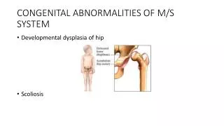
congenital abnormalities
. Let us have a review of the normal anatomy of the urinary system before having a knowledge of the congenital abnormalities. . . . . . . . . . . The most frequent congenital defects and abnormalities of the genitourinary tract are hydronephrosis, undescended testicles (cryptorchidism), hypospadias and epispadias.


congenital abnormalities
E N D
Presentation Transcript
1. Congenital Abnormalities Of the Genitourinary System Dr.V.Kasiviswanathan MS
Formerly Sr.DMO / S.Rly. / Palakkad
13. The most frequent congenital defects and abnormalities of the genitourinary tract are hydronephrosis, undescended testicles (cryptorchidism), hypospadias and epispadias
14. Absence of one kidney Congenital aplasia
Failure to develop one kidney
Can be found during ulatrasound examination, CT scanning and Pyelogram studies
Ureter absent
No ureteric orifice found during cystoscopy
Or ureter and renal pelvis are present but the kidney absent
15. Absent left kidney
16. CT Urography
CT Urography has almost completely replaced conventional excretory urography, popularly called IVU.
With the current multi-slice scanner, especially 64-slice CT scanners, it is possible to obtain high-quality images of the kidneys, ureters and bladder,
Angiographic, parenchymal and excretory phases.
17. Renal Ectopia Kidney does not ascend
Usually near the pelvic brim ; usually left
If it is not symptomatic the only problem is that during the abdominal operations the pelvic kidney should not be mistaken for any abnormal tumour and be injured
18. Renal ectopia � pelvic kidney
19. Horseshoe Kidney Situated usually in front of fouth lumbar vertebra
Fused lower poles common
Ureters angulated
Infection
Nephrolithiasis
Fixed mass below umbilicus
20. Horseshoe Kidney
21. Horseshoe Kidney - pyelogram
22. Horseshoe Kidney � CT scan
23. Horseshoe Scintigram
24. Horse shoe kidney
25. Unilateral Fusion Both kidneys are in one loin
Usually fused
Ureter of the lower kidney crosses the midline to enter the bladder on the contralateral side.
Both renal pelves may lie one above each other medial to the renal parenchyma(unilateral long kidney - or the pelvis of the crossed kidney faces laterally (unilateral S-shaped kidney)
26. Crossed fused kidneys
27. Congenital cystic kidneys � polycystic kidneys Hereditary
Autosomal dominant trait
Not usually detectable until the second or third decades of life and never manifests before the age of 30
Irregular upper quadrant mass
Loin pain
Haematuria
Infection
Hypertension
Uraemia
CT image : multiple cysts in both kidneys
28. Bilateral polycystic kidneys ct
29. Polycystic kidney
30. Simple Renal Cyst Common
Multiple
Diagnosed on ultrasound
Rarely require treatment
Treat only if causing obstruction
31. Renal Cyst
32. Aberrent renal vessels Two or more renal arteries are most common on the left
Functional end arteries � infarction if divided
Veins can be divided because they have collaterals
33. Normal Renal Arteries
34. Two Right Renal Arteries
35. Congenital abnormalities of the renal pelvis and ureter Duplication of a renal pelvis
Common
Usually unilateral
36. Duplication of a ureter
The ureters usually join before they reach the bladder
Less commonly the ureters open indepedently into the bladder
Congenital megaureter
37. Duplication of the ureter
38. Congenital defects of the bladder Ectopia vesicae � exstrophy of the bladder
Easily recognised at birth
Umbilicus absent, protruding due to the intraabdominal pressure
In addition epispadias
Mons and clitoris bifid
In the neonate the bladder should be covered with Saran Wrap or clingfilm to prevent trauma to the delicate mucosa
39. Exstrophy of the bladder
40. Congenital abnormalities of the urethra and penis Meatal stenosis
Congenital stricture
Congenital valves
Hypospadias
epispadias
41. Meatus Congenital stenosis of the external urethral meatus � normally the narrowest part of the male urethra
Associated with phimosis � at times pin hole meatus
Back pressure effects
Spraying, dribbling
42. Congenital Urethral Stricture Rare
43. Congenital valves of the posterior urethra Folds of urothelium
Obstuction in boys
Within prostatic urethra
Catheter will pass easily
Micturating cystourethrogram
Pass catheter
44. Posterior Urethral Valve
45. Micturating cystourethrogram
46. Posterior Urethral Valve with proximal dilatation of the prostatic urethra
47. Hypospadias Most common urethral abnormality
Glandular hypospadias
Coronal hypospadias
Penile and penoscrotal hypospadias
Perineal hypospadias
Avoid circumcision
48. Normal External Urethral Meatus
49. Hypospadias types
50. Hypospadias
51. Abnormalities of the testes and scrotum Incompletely Descended Testis
Testis is not present in the scrotum
In about 4 % of all newborns
50% descend during the first month of life
The genitals all newborns must be examined
May be associated with inguinal hernia
Should be corrected well before puberty
Otherwise atrophies
52. Various Positions of the incompletely descended testis
54. Retractile Testis Sometimes the testis intermittently disappears upwards.
This phenomenon is called 'retractile testis'.
wait for the boy to grow
careful followup
if the testis prefers to stay higher or if the testis is under tension when brought down, surgical correction is recommended.
55. Ectopic testis The sites of ectopic testes are
At the superficial inguinal ring.
In the peruneum
At the root of the penis
In the feroral ring
56. Phimosis At birth foreskin adherent to the surface of the glans penis
Separate spontaneously with time
Can wait for 4 years to separate
Gentle retraction at bath permitted
Forcible retractions injure
57. Phimosis