Bacterial Morphology
This PDF covers bacterial classification based on shape, size, and arrangement u2014 including cocci, bacilli, spirilla, and more. It also explains flagella types, microscopy techniques, and the clinical importance of morphology.<br>ud83eudde0 By Dr. Preeti Tyagi (MBBS,MD) Known as best microbiology faculty in India<br>ud83dudcf2 Full Microbiology Course on Turning Brain App with Unlimited MCQ Practice Test<br>Download the App right now App is available for Android and IOS both.<br>ud83cudf10 www.turningbrain.in | <br>ud83dudcde 91-8368648746<br>You can also watch us on Youtube as @dr.preetityagilectures874


Bacterial Morphology
E N D
Presentation Transcript
Morphology of Bacteria -By Dr. Preeti Tyagi (MBBS,MD) Professor of Physiology VMMC & Sufdarjung Hospital New Delhi
Introduction Bacterial morphology refers to the size, shape, and arrangement of bacterial cells. It's one of the most fundamental aspects of microbiology and is crucial in identifying and classifying bacteria. Understanding morphology helps in: Diagnosing infections Interpreting culture results Studying bacterial evolution The three key features of bacterial morphology include: Shape (form of the cell) Arrangement (how cells group after division) Size (typically 0.2 – 2.0 µm in diameter)
Classification Based on Shape Vibrio (comma-shaped) E.g., Vibrio cholerae Coccus (spherical) E.g., Streptococcus pneumoniae Spirochete (flexible spiral) E.g., Treponema pallidum Bacillus (rod-shaped) E.g., Escherichia coli Spirillum (rigid spiral) E.g., Spirillum minus
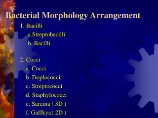
Cocci Arrangement Cocci can appear in various patterns based on their division: Diplococci – in pairs (e.g., Neisseria gonorrhoeae) Streptococci – in chains (e.g., Streptococcus pyogenes) Staphylococci – in grape-like clusters (e.g., Staphylococcus aureus) Tetrads – groups of four (e.g., Micrococcus luteus) Sarcinae – cube of 8 cells (e.g., Sarcina ventriculi)
THANK-YOU Full Physiology Course is available on Turning Brain App by The Best Physiology Faculty in india, Dr. Preeti Tyagi For More Info. +91-8368648746 www.turningbrain.in https://youtube.com/@dr.preetityagilectures874?si=YfbJEi66jOE0hjzu APP LINKS Android: https://play.google.com/store/apps/details id=tbrain.in.medical.eduapp&hl=en_IN IOS: https://apps.apple.com/in/app/turning-brain-dr-preeti-tyagi/id6502828714
Bacilli Arrangement Rod-shaped bacteria (bacilli) have fewer arrangement types: Single bacilli – e.g., Mycobacterium tuberculosis Diplobacilli – two rods (e.g., Coxiella burnetii) Streptobacilli – chains (e.g., Streptobacillus moniliformis) Coccobacilli – short rods resembling cocci (e.g., Haemophilus influenzae)
Other Shaped Bacteria Some bacteria have unique or irregular shapes: Vibrio – curved rod, comma-shaped Spirilla – rigid spiral with flagella Spirochetes – flexible spirals, motile via axial filaments Pleomorphic – variable shape due to lack of rigid cell wall (e.g., Mycoplasma)
Size of Bacteria Bacteria’s are in different sizes: Smallest: Mycoplasma (0.1 µm) Largest: Epulopiscium fishelsoni (up to 750 µm) Most pathogenic bacteria range from 0.5 to 5 µm Size affects nutrient uptake, metabolism, and pathogenicity
Flagella and Motility Flagella help in motility and classification: Monotrichous – one flagellum (e.g., Vibrio cholerae) Lophotrichous – multiple flagella at one end Amphitrichous – flagella at both ends Peritrichous – flagella all over the surface (e.g., E. coli)
Microscopy for Morphology Bacterial morphology can be observed using: Light Microscopy – Basic observation with staining Gram Staining – Differentiates Gram- positive and Gram-negative Electron Microscopy – Detailed 3D surface and internal structures
THANK-YOU Full Physiology Course is available on Turning Brain App by The Best Physiology Faculty in india, Dr. Preeti Tyagi For More Info. +91-8368648746 www.turningbrain.in https://youtube.com/@dr.preetityagilectures874?si=YfbJEi66jOE0hjzu APP LINKS Android: https://play.google.com/store/apps/details id=tbrain.in.medical.eduapp&hl=en_IN IOS: https://apps.apple.com/in/app/turning-brain-dr-preeti-tyagi/id6502828714