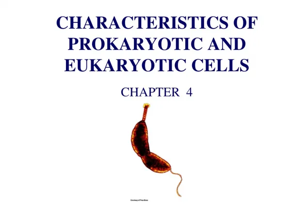
Lecture-3 A Survey of Prokaryotic Cells and Microorganisms
Lecture-3 A Survey of Prokaryotic Cells and Microorganisms. The cell. The cell can be defined : as a basic functional unit of life .

Lecture-3 A Survey of Prokaryotic Cells and Microorganisms
E N D
Presentation Transcript
Lecture-3 A Survey of Prokaryotic Cells and Microorganisms
The cell The cell can be defined : as a basic functional unit of life. The cell was first observed by a scientist named Robert Hook in the 1665. All living organisms are composed of one or many cells to perform their individual functions. All living organisms may composed of one cell (single cell) which called uni-cellular organisms, like prokaryotic organisms including : bacteria, or composed from more than one cell (multi-cellular organisms), like eukaryotic organisms including : human, animals and plants.
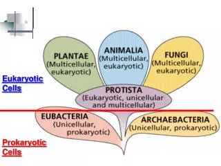
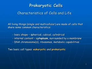
Characteristics of Cells and Life All living things (single and multicellular) are made of cells that share some common characteristics: • Basic shape – spherical, cubical, cylindrical • Internal content – cytoplasm, surrounded by a membrane • DNA chromosome(s), ribosomes, metabolic capabilities Two basic cell types: eukaryotic and prokaryotic
Differences between prokaryotic and eukaryotic cells • -Prokaryotic CellsEukaryotic Cells • ---------------------------------------------------------------------------------------------------- • 1- They are very minute in size. 1-They are larger in size. • 2 -Nuclear region (nucleoid) is not • enveloped by a nuclear membrane. 2- Nucleus is surrounded by a double .. Membrane later • 3- Single chromosome present. 3- More than one chromosome are. • present. • 4- Nucleolus is absent. 4- Nucleolus is present. • 5- Membrane bound organelles are 5- membrane bound organelles are • absent. present. • 6- Multiplication of cell is by fission 6-cell division by mitosis or • or budding. meiosis. • 7-Cell type is usually unicellular. 7-Usually multicellular cells. • 8- Cell size is 1-10μm 8- Cell size 10 - 100µm. • Example: Bacteria, archaea Example: animal cells and plant cells
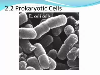
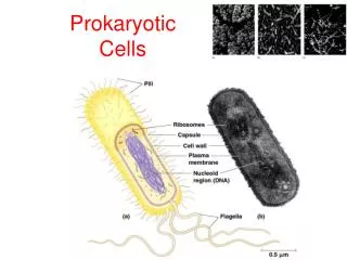
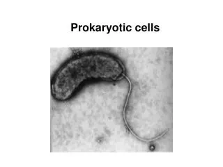
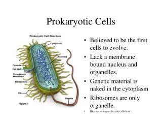
Prokaryotic Cell • Prokaryotic cells are simpler and smaller than the eukaryotic cells. • The term prokaryote is derived from the Greek word- “prokaryote” meaning before nuclei. These cells lack membrane bound organelles. Prokaryotic cells are unicellular organisms, which reproduce through binary fission. In some cases few prokaryotic organisms also reproduce by budding. Prokaryotic cells have a cell envelope, which generally consists of a capsule, cell wall, cytoplasm, plasma membrane, nucleiod region, ribosome, plasmids, pili and flagella
Outer structure Capsule: It is composed of a thick polysaccharide which covers the outside of the cell wall. It is used to stick cells together and works as a food reserve and it also protects the cell from dryness and from chemicals. Cell wall: It is made from the glycoprotein murein. Cell wall provides strength and rigidity to the cell and it is permeable to solutes. Pili: They are short protein appendages, which fixes bacteria to surfaces. These pili are smaller than those flagella and are used in conjugation to exchange the genetic information. Flagella: They are rigid rotating tail. The clockwise rotation moves the cell forward and anticlockwise rotation helps the cell to spin. The rotation is powered by H+ gradient across the cell membrane
External Structures • Appendages • Two major groups of appendages: • Motility – flagella and axial filaments (periplasmic flagella) • Attachment or channels – fimbriae and pili • Glycocalyx – surface coating
Glycocalyx • Coating of molecules external to the cell wall, made of sugars and/or proteins • Two types: • Slime layer - loosely organized and attached • Capsule - highly organized, tightly attached
Functions of the Glycocalyx Protect cells from dehydration and nutrient loss Inhibit killing by white blood cells by phagocytosis, contributing to pathogenicity Attachment - formation of biofilms
Flagella • 3 parts: • Filament – long, thin, helical structure composed of protein flagellin • Hook– curved sheath • Basal body – stack of rings firmly anchored in cell wall
Flagellar Arrangements Monotrichous – single flagellum at one end Lophotrichous – small bunches emerging from the same site Amphitrichous – flagella at both ends of cell Peritrichous – flagella dispersed over surface of cell
FlagellarResponsesflagella Guide bacteria in a direction in response to external stimulus: Chemical stimuli – chemotaxis; positive and negative Light stimuli – phototaxis Signal sets flagella into motion clockwise or counterclockwise: Counterclockwise – results in smooth linear direction – run Clockwise – tumblesf
Fimbriae • Fine, proteinaceous, hairlike bristles emerging from the cell surface • Function in adhesion to other cells and surfaces
Pili • Rigid tubular structure made of pilin protein • Found only in gram-negative cells • Function to join bacterial cells for partial DNA transfer called conjugation
The Cell Envelope • External covering outside the cytoplasm • Composed of two basic layers: • Cell wall and cell membrane • Maintains cell integrity • Two different groups of bacteria demonstrated by Gram stain: • Gram-positive bacteria: thick cell wall composed of- primarily of peptidoglycan and cell membrane • Gram-negative bacteria:cell wall composed of -outer cell membrane, thin peptidoglycan layer, and cell membrane
Structureof Cell Walls and functions • Determines cell shape, prevents lysis due to changing osmotic pressures • Peptidoglycan is the primary component: • Unique macromolecule composed of a repeating framework of long glycan chains cross-linked by short peptide fragments
The Gram Stain • Differential stain that distinguishes cells with a gram-positive cell wall from those with a gram-negative cell wall • Gram-positive - retain crystal violet and stain purple • Gram-negative - lose crystal violet and stain red from safranin counterstain . • Gram stain is: • Important basis of bacterial classification and identification • Practical aid in diagnosing infection and guiding drug treatment
Cell Membrane Structure • Phospholipid bilayer with embedded proteins –this structure called fluid mosaic model • Functions in: • Providing site for energy reactions, nutrient processing, and synthesis • Passage of nutrients into the cell and discharge of wastes • Cell membrane is selectively permeable
Mesosomes • Mesosomes: They are the folding, present inside the plasma membrane. • Their function: Mesosome plays a vital role in cellular respirations, replication of DNA, cell division, separation of chromosomes during cell division and also performs the role of Golgi bodies and mitochondria
Inner structure • Cytoplasm: Cytoplasm is the storehouses for all types of chemicals and components that are used to sustain the life of a bacterium. It helps in cellular growth, metabolism and replication. • 70-80% of cytoplasm is water . • Serves as solvent for materials used in all cell functions
nucleiod region: An area of the cytoplasm that contains the single bacterial DNA molecule. • Plasmids: They are a small circle of DNA. Plasmid plays a vital role in exchanging DNA between the bacterial cells. Bacterial cells have many plasmids. • Chromosome • Single, circular, double-stranded DNA molecule that contains all the genetic information required by a cell
Ribosomes: They are the smallest part of cell organelle. Ribosome plays a vital role in protein synthesis as they consist of protein and RNA. They are located freely in the cytoplasm of attached to the rough endoplasmic reticulum • Prokaryotic differ from eukaryotic ribosomes in size and number of proteins
Internal Structures • Inclusions and granules • Intracellular storage bodies • Vary in size, number, and content • Bacterial cell can use them when environmental sources are depleted
Bacterial Internal Structures • Spores (endospores): Metabolically inert bacterial forms adapted for long-term survival in the environment, which are able to regrow under suitable conditions • produced by some G+ genera: Clostridium, Bacillus, and Sporosarcina • Have a 2-phase life cycle: • Vegetative cell – metabolically active and growing • Endospore – when exposed to adverse environmental conditions; capable of high resistance and very long-term survival
Common Bacterial Shapes, Arrangements, and Sizes • Bacterial cells Vary in shape, size, and arrangement but typically described by one of three basic shapes: • Coccus – spherical • Bacillus – rod • Coccobacillus – very short and plump • Vibrio – gently curved • Spirillum – helical, comma, twisted rod, • Spirochete – spring-like
Pleomorphism • Pleomorphism is:Variation in cell shape and size within a single species • Some species are noted for their pleomorphism
Bacterial Arrangements • Arrangement of cells is dependent on pattern of division and how cells remain attached after division: • Cocci: • Singles • Diplococci – in pairs • Tetrads – groups of four • Irregular clusters • Chains • Cubical packets (sarcina) • Bacilli: • Diplobacilli • Chains • Palisades
Classification Systems for Prokaryotes • We identify microorganisms to predict their pathogenicity. Bacteria are identified using phenotypic, immunological or molecular characteristics that including : • 1- Gram reaction: Gram-positive and Gram-negative bacteria respond to different antibiotics. • 2- Cell shape: Bacteria may be shaped as cocci, bacilli or spirals. • 3- Endospore: The presence, shape and position of the endospore within the bacterial cell are noted
4- Fastidiousness: Certain bacteria have specific O2/CO2 requirements, need special media or grow only intracellularly. • 5- Key enzymes: Some bacteria lack certain enzymes, for example, lack of lactose fermentation helps distinguish salmonellae from E. coli. • 6- Serological reactions: Interaction of antibodies with surface structures may for example help to distinguish subtypes of bacterial species.
Bacterial Taxonomy Based on Bergey’s Manual • Bergey’s Manual of Determinative Bacteriology – five volume resource covering all known prokaryotes • Classification based on genetic information –phylogenetic • Two domains: Archaea and Bacteria • Five major subgroups with 25 different phyla
Major Taxonomic Groups of Prokaryotes • Domain Archaea – primitive, adapted to extreme habitats and modes of nutrition • Domain Bacteria: • Phylum Proteobacteria – Gram-negative cell walls • Phylum Firmicutes – mainly Gram-positive with low G + C content • Phylum Actinobacteria – Gram-positive with high G + C content
Species and Subspecies • Species – a collection of bacterial cells which share an overall similar pattern of traits in contrast to other bacteria whose pattern differs significantly • Strain or variety – a culture derived from a single parent that differs in structure or metabolism from other cultures of that species (biovars, morphovars) • Type – a subspecies that can show differences in antigenic makeup (serotype or serovar), susceptibility to bacterial viruses (phage type) and in pathogenicity (pathotype)
Prokaryotes with Unusual Characteristics • Free-living nonpathogenic bacteria • Photosynthetic bacteria – use photosynthesis, can synthesize required nutrients from inorganic compounds • Cyanobacteria (blue-green algae) • Green and purple sulfur bacteria • Gliding, fruiting bacteria
Unusual Forms of Medically Significant Bacteria • Obligate intracellular parasites • Rickettsias • Very tiny, gram-negative bacteria • Most are pathogens • Obligate intracellular pathogens • Cannot survive or multiply outside of a host cell • Rickettsia rickettisii – cause :Rocky Mountain spotted fever
Unusual Forms of Medically Significant Bacteria • Chlamydias • Tiny cell • Obligate intracellular parasites • Not transmitted by arthropods • Chlamydia trachomatis – cause severe eye infection and one of the most common sexually transmitted diseases • Chlamydia pneumoniae – cause lung infections
Archaea Constitute third Domain Archaea More closely related to Eukarya than to Bacteria Contain unique genetic sequences in their rRNA Have unique membrane lipids and cell walls
Archaea Live in the most extreme habitats in nature, extremophiles Adapted to heat, salt, acid pH, pressure, and atmosphere Includes: methane producers, hyperthermophiles, extreme halophiles, and sulfur reducers 39 5 mm