UTERINE FIBROID
UTERINE FIBROID. أهلا بحضراتكم. التعارف. 6 th Y. Anticipations. Learning Objectives. LEARNING OBJECTIVES Describe the pathology of uterine fibroids List complications of uterine fibroids Describe symptoms and signs of fibroids Interpret different investigations and DD of fibroids

UTERINE FIBROID
E N D
Presentation Transcript
أهلا بحضراتكم إدارة التدريب
التعارف 6th Y
LEARNING OBJECTIVES • Describe the pathology of uterine fibroids • List complications of uterine fibroids • Describe symptoms and signs of fibroids • Interpret different investigations and DD of fibroids • Describe different lines of treatment according to different types and conditions • State complications of fibroids during pregnancy • List types of uterine polyps
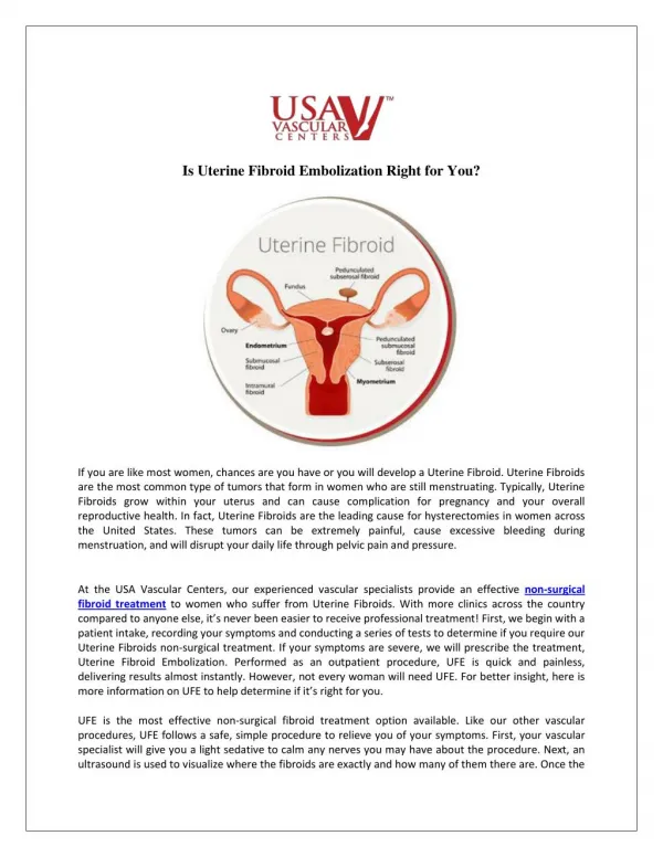
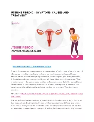
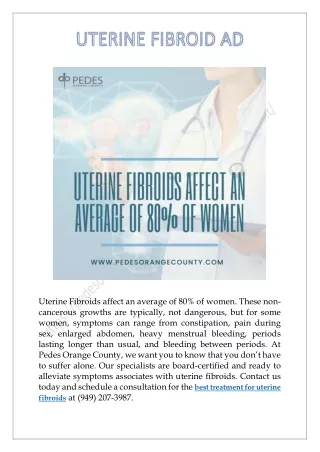
Definition. it is a benign tumor of the smooth muscle of the uterus. Incidence. Fibroids are the commonest tumors of the uterus occurring mostly in patients about 35-40 years who are usually nulliparas or of low parity. It is probable that relative estrogen excess is a predisposing factor.
Etiology The exact etiology is unknown . however the following factor are predisposing • Age. • Parity. • Race: more in black. • Family history. • Hormonal influences: relative estrogen excess.
PATHOLOGY Gross: • Uterine fibroids starts interstitial (intramural) but may become submucous or subserous (the last 2 may become pedunculated) and subserous fibroid may grow between the 2 layers of the broad ligament. • surrounded by a pseudocapsule (false capsule) composed of compressed normal uterine muscles.
2ry PATHOLOGICAL CHANGES OCCURRING IN FIBROIDS: I-degenerative changes • Atrophy. • Necrosis. • Hyaline degeneration. • Cystic degeneration. • Red degeneration. • Fatty degeneration. • Calcification. • Malignant change. II-vascular changes. III-infection.
Complications of Fibroid • Torsion of a pedunculated subserous fibroid. • Rupture of a surface vein of subserous fibroid causing internal hemorrhage. • Infection. • Red degeneration. • Malignant change. • Chronic inversion of the uterus in cases of submucous fundal fibroid. • Impaction of the fibroid (incarceration). This may occur with cervical fibroid
Symptoms of fibroid • Asymptomatic: accidentally discovered • Menorrhagia. • Metrorrhagia. • Congestive dysmenorrhoea. • Leukorrhoea. • Infertility. • Abdominal mass. • Pain. • Pressure symptoms.
Investigations • Ultrasonography. • Hysterosalpingography. • Intravenous pyelography. • MRI • Dilatation and curettage. In case of metrorrhagia. • Routine investigation to prepare the patient to operation.
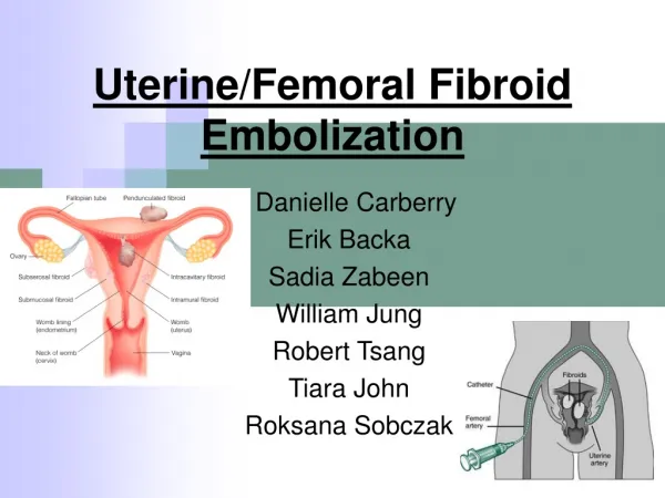
CLASSIFICATION • Submucous leiomyoma • Pedunculated submucous • Intramural or interstitial • Subserous or subperitoneal • Pedunculated abdominal • Parasitic • Intraligmentary • Cervical
Differential Diagnosis • Other causes of pelviabdominal mass. • Other causes of menorrhagia. • Other causes of mass protruding from the cervix: in case of submucous fibroid.
Treatment of fibroid. The treatment depends on: • Age and parity. • Number, size and types of the tumors. • Severity of symptoms. • Complications especially suspicion of malignancy. • Associated pathological changes as cancer body . • Associated pregnancy.
I- No treatment. Small symptomless fibroids especially near menopause require no treatment but the patient should be kept under observation. II-surgical treatment. Myomectomy or hysterectomy. III-other line of treatment.
Myomectomy • It consists of shelling out the tumor from the uterus (ppt) followed by obliteration of the cavities left behind (ppt). It is done usually abdominally but occasionally vaginally .
Contraindications of Myomectomy • Patients near menopause. • Very large number of fibroid so that the uterus left will be useless. • Suspicion of malignancy. • Large interstitial cervical fibroid, fixed into the pelvis.
Disadvantages of myomectomy • Higher mortality than hysterectomy. • Persistence of menorrhagia. • Recurrence. • Pelvic adhesions. • Rupture of the scar may occur in subsequent pregnancy.
Cervicalpolyps • Mucous polyp. • Fibroadenomatous polyp. • Fibroid polyp. • Malignant polyp. • Belharzial papilloma.
Uterine polypi A-Corporeal polypi. • Adenomatous polyp. • Fibroid polyp. • Placental polyp. • Malignant polyp.
Did you meet your anticipation? إدارة التدريب
عزيزي الطالب إدارة التدريب يرجي الانتباه لضرورة الدخول على موقع الكلية: http://www.sohag-univ.edu.eg/facemed/ إدارة التدريب
Reflection- Seminar- Portfolio إدارة التدريب
Thank you يسعدنا التواصل مع حضراتكم http://www.sohag-univ.edu.eg/facemed/ 01001986936 Mostafa_atya@med.sohag.edu.eg 30 إدارة التدريب