Uterine Cervix
Uterine Cervix. Emad Raddaoui , MD, FCAP, FASC. SQUAMO-COLUMNAR JUNCTION. Erosion/Ectropion. Characterized by columnar epithelium replacing squamous epithelium, grossly resulting in an erythematous area.

Uterine Cervix
E N D
Presentation Transcript
Uterine Cervix EmadRaddaoui, MD, FCAP, FASC
Erosion/Ectropion • Characterized by columnar epithelium replacing squamous epithelium, grossly resulting in an erythematous area. • It is a typical response to a variety of stimuli including hormones, chronic irritation and inflammation (chronic cervicitis). • It is benign and has no malignant potential.
Cervical polyp • This is a small, pedunculated, often sessile mass. • Most originate from the endocervix (endocervical polyps) with a few from the ectocervix (ectocervical polyps). • They are inflammatory proliferations of cervical mucosa and are not true neoplasms. • The lesion is characterized by overgrowth of benign stroma covered by epithelium. • Endocervical polyps are covered by endocervical, squamo-columnar or metaplastic squamous epithelium while ectocervical ones are covered by stratified squamous epithelium. • The stroma contains thick-walled blood vessels and fibrous and some inflammatory cells.
CERVICITIS • Inflammation of cervix. • Can be non-infectious or infectious.
Noninfectious (Nonspecific) Cervicitis • This is inflammation of the cervix caused by chemical (e.g. douche, deodorant) or mechanical (e.g. tampon, diaphragm) irritation. It is often acute but may be chronic. Clinical appearances • Noninfectious cervicitis is often asymptomatic. • The cervix appears red and swollen Histology • The histologic features are nonspecific. The inflammatory infiltrate may comprise neutrophils or plasma cells and lymphocytes or a combination of these cells. • Squamous metaplasia of the endocervical glandular epithelium is common in chronic cervicitis. Often some of the mucous glands are obstructed and dilate to form mucus-filled cysts called nabothian cysts.
Infectious cervicitis • Can be caused by various organisims e.g.staphylococci, enterococci, Gardnerella vaginalis, Trichomonas vaginalis, Candida albicans and Chlamydia trachomatis. • Most often involves the endocervix. • Is often asymptomatic • may manifest as vaginal discharge
Candidiasis (moniliasis) • Common form of vaginitis /cervicitis. • Caused by Candida albicans, a normal component of the vaginal flora. • Associated with diabetes mellitis, pregnancy, broad spectrum antibiotic therapy, oral contraceptive use and immunosupression. • Characterized by white patchy mucosal lesions with thick white discharge and vulvovaginalpruritis. Histology. • Colonies of the fungus are present on the surface and extend into the epithelium but not into the underlying tissues. Mild edema and chronic inflammatory cells are present. Ulcers may develop. Cytological smears show yeast forms and branching pseudohyphae
Trichomoniasis • caused by a unicellular flagellated protozoan, Trichomonasvaginalis. It is sexually transmitted disease • Involves the vagina and cervix also. Clinical presentation • Most infections are asymptomatic and pass unnoticed. Occasionally, a copious, thin, frothy, yellow green to gray offensive discharge is produced. There may be vulvar itching or burning or dyspareunia Histology • An inflammatory infiltrate of lymphocytes and plasma cells. The organisms are not seen in biopsies because they do not invade the vaginal wall. Diagnosis is made by examination of a saline wet preparation in which the motile trophozoitesare seen. The organism can also be found in Pap-stained vaginal smears.
Chlamydia trachomatisCervicitis • Clamydiatrachomatis is an obligate, gram-negative intracellular pathogen. • Clamydialcervicitis is the most common sexually transmitted disease in the developed countries. It may coexist with Neisseriagonorrhoeae infection. • It is a frequent cause of pelvic inflammatory disease. • Is most often asymptomatic. • Chlamdial infection may also cause a condition known as lymphogranulomavenereum Clinical appearances • The disease may be symptomatic or asymptomatic. In symptomatic cases there is a mucopurulent cervical discharge with a reddened, congested and edematous cervix. It may be associated with urethritis.
Herpes simplex virus (HSV) Cervicitis • HSV Type 2 infection accounts for majority of genital herpes cases and is spread by sexual contact. • It produces vesicles and ulcers that can involve the cervix, vagina, vulva, urethra and perianal skin.
Human papilloma virus(HPV) infection • HPV infection of the cervix is common. • Over 20 serotypes infect the female genital areas. They cause a variety of different lesions with the different serotypes. Clinical behavior • HPV infection is associated with increased risk of subsequent cervical cancer and so long-term follow-up with attention to the cervix, vagina and vulva is necessary. HPV infection may cause any of the following depending on the serotype 1) Condyloma. This develops in the squamous epithelium of the ectocervix and in foci of squamous metaplasia in the endocervix. The lesions may be flat or exophyticcondylommaacuminatum. Can be caused by any HPV serotype but more commonly by types 6 and 11. 2) Mild dysplasia is usually caused by "low risk" HPV serotypes, 6 and 11. 3) High- grade dysplasia is caused by "high risk” HPV (types16 and 18) and moderate risk HPV( types 31,33,35).
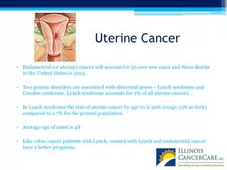
Cervix Carcinoma • One of the major causes of cancer-related death in women, specially in developing world. • Most common cervical cancer is squamous cell carcinoma. Other types are adenocarcinoma, neuroendocrine carcinoma etc. • Nowadays there is dramatic improvement because of early diagnosis and treatment. • The wide use of PAP screening lowered the incidence of invasive cancer .
Precancerous lesion of cervical carcinoma (CIN/ SIL) • All invasive squamous cell carcinomas arise from pre-cancer (non invasive lesions) epithelial changes referred as Cervical Intraepithelial Neoplasia (CIN ) or Squamous intraepithelial lesions. • Squamous Intraepithelial Lesion (SIL) is the terminology used in cytology and CIN is the terminology used in histology(biopsies) • Timely detection and diagnosis of CIN is essential in preventing the development of carcinoma (invasive lesion) and therefore providing curative treatment possible. • Not all cases of CIN progress to invasive cancer and some cases of CIN may spontaneously regress. • The risk of persistence or progression to cancer is more in the high grade precancerous lesions which are associated the high-risk HPV types.
Cervical intraepithelial neoplasia (CIN) • Cytologic examination can detect CIN (SIL) long before any abnormality can be seen grossly. • Pre-cancer changes can precede the development of an overt cancer by many years. • CIN lesions may begin as Low Grade CIN and progress to High Grade CIN, or they might start as HG lesion. • On the basis of histology, pre-cancer lesions are graded as follows: -CIN I : Mild Dysplasia -CIN II : Moderate Dysplasia -CIN III : Severe Dysplasia and Carcinoma in situ.
Downloaded from: Robbins & Cotran Pathologic Basis of Disease
HPV infection • illustrating: • Flat condyloma with prominent koilocytoticatypia of HPV in the upper epithelial cells, as evidenced by the prominent perinuclear halos. • Nucleic acid in situ hybridization of the same lesion for HPV nucleic acids. The dark staining denotes HPV DNA, which is typically most abundant in the koilocytes. • Diffuse immunostaining of CIN II for Ki-67, illustrating widespread deregulation of cell cycle controls. • Up-regulation of p16ink4 (seen as intense immunostaining) characterizes high-risk HPV infections.
CIN I • CIN I with HPV associated koilocytotic atypia • Lower 1/3rd of the epithelium is replaced by pleomorphic cells .
CIN II • CIN II with progressive atypia in the layers of the epithelium; • Lower 2/3rd of the epithelium is replaced by pleomorphic cells
CIN III/ carcinoma in situ • CIN III (carcinoma in situ) with diffuse atypia and loss of maturation • All levels of the epithelium are replaced by pleomorphic cells, (full thickness)
Cytology screening for precancerous lesions • Cytologic examination can detect precancerous lesions long before any abnormality can be seen grossly • For cytologic examination the cervix is examined and the cells lining the cervical wall at the transformation zone are scrapped/ sampled with a spatula and then spread on a slide. They are then fixed, stained (Papanicolaou stain) and examined under a light microscope. • This screening for precancer should be done on all young and old women (usually from age of 21 onwards).
Cytology Pap Smear/Screening In cytology smears we separate pre-cancer lesions into two groups : • Low Grade SIL (= CIN1/mild dysplasia) • High Grade SIL (= CIN2 and 3/moderate to severe dysplasia) • Of Low Grade SIL 1-5 % become invasive • Of High Grade SIL incidence is 6-74%
The cytology of cervical intraepithelial neoplasia as seen on the Papanicolaou smear. Cytoplasmic staining in superficial cells (A&B) may be either red or blue. A, Normal exfoliated superficial squamous epithelial cells. B, CIN I/ low grade SIL C, CIN II/ high grade SIL. D, CIN III/ high grade SIL. Note the reduction in cytoplasm and the increase in the nucleus to cytoplasm ratio, which occurs as the grade of the lesion increases. This reflects the progressive loss of cellular differentiation on the surface of the lesions from which these cells are exfoliated.
CIN/ SIL: Risk Factors • Early age at first intercourse • Multiple sexual partners • A male partner with multiple previous sexual partners • Persistent infection by high risk papillomaviruses • Other risk factors; low socioeconomic groups • rare among virgins, multiple pregnancies. Causes • The cause is determined to be HPV virus .The HPV is the number one reason for abnormal cells of the cervix. • HPV is a skin virus, which results in warts, common warts ,flat warts, genital warts (condylomas), planter warts, and precancerous lesions. • HPV can be detected in 85 -90 % of pre-cancer lesions. • High risk types HPV : 16, 18, 31, 33, 35, 39, 45, 52, 56, 58, and 59. • Low risk types HPV :6, 11, 42, 44 . These types result in condylomas. Treatment • laser or cone biopsy is the most effective method of managing patients with High grade SIL in cancer prevention
Cervical carcinoma, Signs of CIN/ SIL • There are no visible symptoms that you have dysplasia of the cervix, an is difficult to diagnose without a Pap smear or Pap exam . • This is why we should have regular pap exams, as to detect any abnormal cells. Screening • The Pap smear detects early HPV infection. • The common testing procedure for HPV infection is pap exam. Should start at the age 21. • Screening between age 21-29: Cytology screening test should be done every 3 years • There is a HPV DNA in-situ hybridization (ISH) test, called the DiegeneHyprid Capture test to identify the viral strain. This test will determine whether you carry high or low risk strains of the virus. • DNA screening test should not be used before age 30. • Screening between age30-64 : screening by Cytology + HPV test every 5 years (cytology every 3 years is acceptable).
Cervical Carcinoma, Invasive • 75 -90% of invasive cancers are Squamous cell carcinomas ,which generally evolve from pre-cancer CIN. • The remainder are Adenocarcinoma. • Squamous cell cancers are appearing in increasingly younger women, now with a peak incidence at about 45 years, about 10-15 years after detection of their precursors.
Cervical Carcinoma (invasive carcinoma), Morphology • Mainly in the region of the transformation zone, and range from microscopic foci of early stromal invasion to grossly frank tumors encircling the cervical Os . • The tumors may be invisible or exophytic . • Cervical carcinomas are graded from 1 to 3( i.e. well, moderately and poorly differentiated) based on cellular differentiation and staged from 1 to 4 depending on clinical spread.
Cervical Carcinoma, Staging 0- Carcinoma in Situ 1- Confined to the cervix 2- Extension beyond the cervix without extension to the lower third of Vagina or Pelvic Wall 3- Extension to the pelvic wall and / or lower third of the vagina 4- Extends to adjacent organs
Cervical Carcinoma, Clinical Course • Many of cervical cancers are diagnosed in early stages, and the vast majority are diagnosed in the pre-invasive phase. • More advanced cases are seen in women who either have never had a Pap smear or have waited many years since the prior smear. • The early stages of cervical cancer may be completely asymptomatic. • Vaginal bleeding, contact bleeding, or (rarely) a vaginal mass may indicate the presence of malignancy. Also, moderate pain during sexual intercourse and vaginal discharge are symptoms of cervical cancer. In advanced disease, metastases may be present in the abdomen, lungs or elsewhere. • Symptoms of advanced cervical cancer may include: loss of appetite, weight loss, fatigue, pelvic pain, back pain, leg pain, swollen legs, heavy bleeding from the vagina, bone fractures, and/or (rarely) leakage of urine or faeces from the vagina
Cervical Carcinoma: Treatment • Depending on the stage the treatment options included are • If you still want to be able to have children, first the cancer is removed with a cone biopsy (cervical conization), and then you are watched closely to see if the cancer comes back. • Simple hysterectomy (removal of the whole uterus including part of the vagina). • Radical hysterectomy (removal of the whole uterus including part of the vagina along with the removal of lymph nodes in the pelvis. • Adjunct chemotherapy and radiotherapy.
A, Carcinoma of the cervix, well advanced. B, Early stromal invasion occurring in a cervical intraepithelial neoplasm