Download
1 / 2
0 likes | 19 Vues
Immunohistochemistry (IHC) has revolutionized cancer research by enabling the visualization and characterization of specific proteins within tissue samples. This protocol serves as a guide for conducting successful IHC experiments, providing valuable insights into cancer biology.

E N D
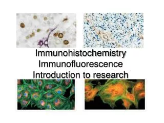
Unlocking Insights: A Comprehensive Immunohistochemistry Protocol for Cancer Research Immunohistochemistry (IHC) has revolutionized cancer research by enabling the visualization and characterization of specific proteins within tissue samples. This protocol serves as a guide for conducting successful IHC experiments, providing valuable insights into cancer biology. 1.Sample Preparation: Begin by obtaining tissue samples from the target area of interest. Ensure proper fixation and embedding to maintain tissue integrity throughout the staining process. 2.Antigen Retrieval: Treat tissue sections with appropriate antigen retrieval solutions to unmask target antigens and enhance antibody binding. Heat-induced epitope retrieval (HIER) methods are commonly employed for this purpose. 3.Blocking: Block nonspecific binding sites by incubating tissue sections with a blocking agent such as serum or protein blockers. This step minimizes background staining and enhances specific antibody binding. 4.Primary Antibody Incubation: Apply the primary antibody specific to the target protein of interest. Incubate the tissue sections overnight at optimal conditions to allow for antibody-antigen binding. 5.Washing: Thoroughly wash tissue sections to remove unbound primary antibodies and excess blocking reagents. Proper washing steps are crucial for reducing background noise and improving signal-to-noise ratios. 6.Secondary Antibody Incubation: Apply a secondary antibody conjugated to a detection molecule (e.g., HRP, AP) that recognizes the primary antibody. Incubate tissue sections under optimal conditions to facilitate secondary antibody binding. 7.Signal Detection: Visualize antibody binding by applying a chromogenic substrate or fluorescent dye that reacts with the detection molecule. Monitor staining intensity and adjust incubation times as needed to optimize signal detection. 8.Counterstaining: Optionally, counterstain tissue sections with a contrasting dye to enhance tissue morphology and aid in sample interpretation. Hematoxylin and eosin (H&E) staining is commonly used for this purpose. 9.Mounting and Imaging: Mount stained tissue sections onto glass slides using an appropriate mounting medium. Capture high-resolution images using a microscope equipped with brightfield or fluorescence capabilities.
10.Data Analysis: Analyze staining patterns and intensities to evaluate protein expression levels and localization within the tissue samples. Quantitative image analysis software can aid in objective data interpretation. By following this comprehensive immunohistochemistry protocol, researchers can unlock valuable insights into cancer biology, including protein expression patterns, cellular localization, and biomarker identification. These insights contribute to the development of targeted therapies and personalized treatment strategies, ultimately advancing the fight against cancer. Address : Unit D, 3/F., Freder Centre Mok Cheong Street, Tokwawan, Hong Kong Ph No : 13808832613 Email : info@ihc-prs.com Website : https://ihc-prs.com/