Download
1 / 2
20 likes | 38 Vues
With the improvement of cutting edge NGS-based oncology quality boards, it is winding up progressively essential to consider pre-logical factors while extricating nucleic acids from FFPE-treated examples. Visit https://immunostaining.info

E N D
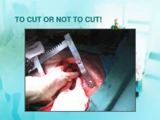
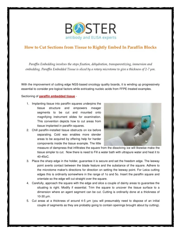
How to Cut Sections from Tissue to Rightly Embed In Paraffin Blocks Paraffin Embedding involves the steps fixation, dehydration, transparentizing, immersion and embedding. Paraffin Embedded Tissue is sliced by a rotary microtome to give a thickness of 2-7 μm. With the improvement of cutting edge NGS-based oncology quality boards, it is winding up progressively essential to consider pre-logical factors while extricating nucleic acids from FFPE-treated examples. Sectioning of paraffin embedded tissue - 1. Implanting tissue into paraffin squares underpins the tissue structure segments to be cut and mounted onto magnifying instrument slides for examination. This convention depicts how to cut areas from tissue implanted in paraffin squares. 2. Chill paraffin-installed tissue obstructs on ice before separating. Cold wax enables more slender areas to be acquired by offering help for harder components inside the tissue example. The little measure of dampness that infiltrates the square from the dissolving ice will likewise make the tissue simpler to cut. Now there is need to Fill a water bath with ultrapure water and heat it to 40-45oC. 3. Place the sharp edge in the holder, guarantee it is secure and set the freedom edge. The leeway point averts contact between the blade feature and the substance of the square. Adhere to the microtome maker's directions for direction on setting the leeway point. For Leica cutting edges this is ordinarily somewhere in the range of 1o and 5o. Insert the paraffin square and orientate so the edge will cut straight over the square. 4. Carefully, approach the square with the edge and slice a couple of dainty areas to guarantee the situating is right. Modify if essential. Trim the square to uncover the tissue surface to a dimension where an agent segment can be cut. Cutting is ordinarily done at a thickness of 10-30 µm. 5. Cut areas at a thickness of around 4-5 µm (you will presumably need to dispose of an initial couple of segments as they are probably going to contain openings brought about by cutting). and empowers meager
Using tweezers get the strips of segments and buoy them on the outside of the water in the water shower so they level out. Utilize the tweezers to isolate the areas. 6. Use magnifying instrument slides to choose segments from the water shower and store upstanding in a slide rack. Place the slide rack into a stove and enable segments to successfully dry medium-term at 37oC. Your quest for getting great guidance about Tissue Sectioning completes with Boster Antibody and Elisa Experts. Get assistance from veteran experts https://immunostaining.info/ from the web portal. Visit the link Contact Us Immunohistochemical Staining 3942 Valley Ave Pleasanton, CA 94566, USA Email:support@bosterbio.com Phone: (888) 466-3604