Understanding ICC Protocol Immunohistochemical Staining
20 likes | 37 Vues
ICC Protocol Immunohistochemical Staining has turned into a routinely utilized approach for insightful and indicative assessments. visit https://immunostaining.info

Understanding ICC Protocol Immunohistochemical Staining
E N D
Presentation Transcript
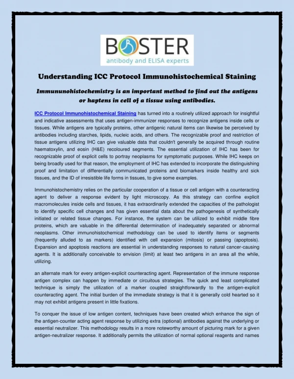
ICC Protocol Immunohistochemical Staining has turned into a routinely utilized approach for insightful and indicative assessments that uses antigen-immunizer responses to recognize antigens inside cells or tissues. While antigens are typically proteins, other antigenic natural items can likewise be perceived by antibodies including starches, lipids, nucleic acids, and others. The recognizable proof and restriction of tissue antigens utilizing IHC can give valuable data that couldn't generally be acquired through routine haematoxylin, and eosin (H&E) recoloured segments. The essential utilization of IHC has been for recognizable proof of explicit cells to portray neoplasms for symptomatic purposes. While IHC keeps on being broadly used for that reason, the employment of IHC has extended to incorporate the distinguishing proof and limitation of differentially communicated proteins and biomarkers inside healthy and sick tissues, and the ID of irresistible life forms in tissues, to give some examples. Immunohistochemistry relies on the particular cooperation of a tissue or cell antigen with a counteracting agent to deliver a response evident by light microscopy. As this strategy can confine explicit macromolecules inside cells and tissues, it has extraordinarily extended the capacities of the pathologist to identify specific cell changes and has given essential data about the pathogenesis of synthetically initiated or related tissue changes. For instance, the system can be utilized to exhibit middle fibre proteins, which are valuable in the differential determination of inadequately separated or abnormal neoplasms. Other immunohistochemical methodology can be used to identify items or segments (frequently alluded to as markers) identified with cell expansion (mitosis) or passing (apoptosis). Expansion and apoptosis reactions are essential in understanding responses to natural cancer-causing agents. It is additionally conceivable to envision (limit) at least two antigens in an area all the while, utilizing. an alternate mark for every antigen-explicit counteracting agent. Representation of the immune response antigen complex can happen by immediate or circuitous strategies. The quick and least complicated technique is simply the utilization of a marker coupled straightforwardly to the antigen-explicit counteracting agent. The initial burden of the immediate strategy is that it is generally cold hearted so it may not exhibit antigens present in little fixations. To conquer the issue of low antigen content, techniques have been created which enhance the sign of the antigen-counter acting agent response by utilizing extra (optional) antibodies against the underlying or essential neutralizer. This methodology results in a more noteworthy amount of picturing mark for a given antigen-neutralizer response. It additionally permits the utilization of normal optional reagents and names
for various essential antibodies. The avidin-biotin strategy is one case of this kind of aberrant enhanced technique. Markers extend from fluorescent colours to thick materials, for example, colloidal gold. The most widely recognized IHC methods use new advances, for example, chemical response in the creation of a noticeable chromogen (e.g., peroxidase response procedures) or a strengthening framework utilizing photographic designers (e.g., colloidal gold strategies). The immunogold-silver recolouring strategy has been demonstrated to be of significant worth in the identification of administrative peptides in nerves and endocrine cells in routinely fixed (formalin fixed), paraffin implanted tissues. About Boster Bio As the leading Flow Cytometry Technical Resource Centers, Boster Bio was founded by Steven Xia, histologist, in the year 1993. The business currently provides hybridoma generation check to product custom monoclonal antibodies. Find ICC Protocol Immunohistochemical Staining that covers procedure included in cell climbing slice preparation. For more information on Paraffin Embedded Tissue, please visit https://immunostaining.info Contact Address Boster Biological Technology 3942 Valley Ave Pleasanton, CA 94566, USA Email:support@bosterbio.com Phone: (888) 466-3604