Download

1 / 26
260 likes | 574 Vues
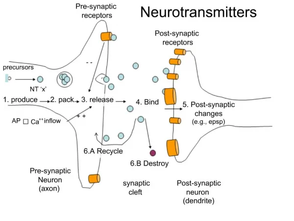
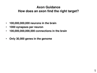
Axon guidance. Neuronal growth cones respond to both contact mediated and chemotropic guidance cues. Cues can be either attractive or repulsive. Cues can act over a short or long range, and may differentially affect particular types of neurons. Only the growth cone is motile.

E N D
Neuronal growth cones respond to both contact mediated and chemotropic guidance cues. • Cues can be either attractive or repulsive. • Cues can act over a short or long range, and may differentially affect particular types of neurons. • Only the growth cone is motile
Guidance by adhesive gradients: Haptotaxis • Growth cone expresses cell adhesion molecules (CAMs), enable it to move by modulating adhesion to the extracellular matrix and other cells. • CAMs on growth cone recognise proteins found in certain basal laminae • Laminin appears to pave many axonal tracts, even if only transiently. • Glycosaminoglycans appear to impede neural outgrowths • Because laminin and N-CAM are found in several places, they can only provide general cues.
Four conserved families of guidance cues • netrins • slits • semaphorins • ephrins • Netrins, slits and some semaphorins are secreted and associate with cells or the ECM • Ephrins and some semaphorins are membrane bound • netrins can act as attractants or repellants • slits, semaphorins and ephrins act primarily as repellants but can be attractant in some cases • for each cue there is one or more transmembrane receptor
Netrins • Netrins are a small family of highly conserved guidance molecules (~70-80kDa). • One in worms (c.elegans) UNC6 • Two in Drosophila Netrin -A and -B • Two in chick, netrin-1 and -2. • In mouse and humans a third netrin identified netrin-3 (netrin-2-like). • In all species there are axons that project to the midline of the nervous system. • The midline attracts these axons and netrin plays a role in this.
Netrin-1 is produced by the floor plate • Netrin-2 is made in the ventral spinal cord except for the floor-plate • Both netrins become associated with the ECM and the receptor DCC • Model: commissural axons first encounter gradient of netrin-2, which brings them into the domain of netrin-1
Roof plate Commissural neuron 0 125 250 375 Commissural neurons extend ventrally and then toward floor plate, if within 250mm from the floor plate Floor plate
Netrins are bifunctional molecules, attracting some axons and repelling others. • C.elegans axons migrating away from the UNC-6 netrin source are misrouted in the unc-6 mutant. • The repulsive activity of netrin first shown in vertebrates for populations of motor axons that project away from the midline. • The receptors that mediate the attractive and repulsive effects of netrins are also highly conserved. • Growth cone attraction involves the transmembrane receptors of the DCC family. • Repulsion involves the transmembrane receptors of the UNC-5 family.
Netrin=unc6 Ligand DCC=unc40 attract unc5 = repulsion Unc-6 minus Wild type Unc-40+ sensory neuons Unc-5+ motor neurons UNC-6 Unc-5 minus Unc-40 minus
Slits • Slit proteins are large ~190kDa extracellular matrix proteins containing leucine-rich repeats and epidermal growth factor-like repeats. • Slit has a role in axon guidance at the midline. • Receptor (Robo) is expressed by axons that navigate the midline and prevents them crossing • Robo expressing axons run longitudinally and never cross the midline. • In Robo mutants axons freely cross the midline. • Commissural axons up-regulate Robo after they have crossed the midline • Slit is repellent ligand
3 vertebrate slit proteins are expressed at the midline and have chemorepulsive activity for olfactory bulb axons, hippocampal axons and spinal motor axons in culture
Semaphorins • First semaphorins identified in grasshopper (Sema-1a/fasciclin IV) and chicken (Sema3a/collapsin-1/Sema III). • Family contains over 20 members. • All characterised by the presence of a ~500 amino acid semaphorin motif in their extracellular domain. • Insects and worms have two sub-families: transmembrane (class 1) and secreted (class 2). • Vertebrates have at least 5 sub-families. • Best characterised function of semaphorins is in axon repulsion.
Secreted Sema3A (Collapsin-1/Sema III) is a potent growth cone collapser, and can repel axons when presented. • Other secreted semaphorins (vertebrate class 3) also repulse axons. • Because of the large number of family members differential effects are seen for axon repulsion. • Sema3C acts an attractant signal for cortical axons • Sema IV has a strong repulsive action on, olfactory axons, whereas Sema A appears to attract.
2 distinct classes of semaphorin receptors identified. • In vertebrates the transmembrane proteins neuropilin-1 and -2 act as receptors required for mediating the repulsive actions of class 3 semaphorins • Sema3A signal is mediated by neuropilin-1 • Sema3F by neuropilin-2 • Sema3C by neuropilin-1/-2 heterodimers. • Neuropilins are only found in vertebrates in flies plexin A mediates repulsion by Sema-1A in Drosophila • Plexins have also been identified in vertebrates.
Ephrins and eph’s Eph receptors and ephrins are a major class of contact-dependent repellants in vitro theycan trigger growth-cone collapse and inhibit neurite outgrowth. In vivo they can channel axons along specific routes by preventing growth cones from migrating across their expression boundaries;
Include navigation of commissural axons across the midline of the forebrain and midbrain, • Restriction of axons to longitudinal tracts that do not cross the ventral midline of the spinal cord, • Pathfinding of spinal motor axons through only the anterior half of somites and pathfinding of motor axons in the limb. • In frogs, even if the eye is inverted through 180o the axons still find their original sites of contact. • Suggests that each retinal neuron carries a chemical label that enables it to connect reliably with an appropriately chemically labelled cell in the tectum.
Different situations in which Eph receptors and ephrins restrict cell or growth-cone migration by repulsion at boundaries. a Bidirectional activation of Eph receptor and ephrin-B ligand restricts mixing between adjacent cell populations by mediating mutual repulsion. b Boundaries of ephrin expression repel migrating cells expressing Eph receptor, so restricting them to adjacent territories. c Boundaries of ephrin expression restrict growth cones of neurons expressing Eph receptor. d Boundaries of Eph receptor restrict growth cones of neurons expressing ephrin-B protein. Directional activation of Eph receptors and ephrins indicated by blue and red arrows.
a In retinotectal topographic map, axons project from temporal retina to anterior tectum, and from nasal retina to posterior tectum. b A graded sensitivity of retinal axons to ephrin-A2 and ephrin-A5, repellents expressed in overlapping gradients in the tectum. The graded sensitivity of axons is due to graded expression of EphA3 or EphA5, and to overlapping expression of uniform EphA4 (blue line) and graded ephrin-A5 (red line) expression, which underlies graded sensitivity of EphA4 along the temporal/nasal axis (black dotted line).
alternating posterior and anterior tectum membranes show that: • Nasal retinal cells extended axons equally well on both anterior and posterior tectal membranes. • Temporal side retinal cells only extended axons on the anterior tectal membranes. • Growth cones that extend on the posterior membranes collapse and retract. • EphA receptors and ephrinA ligands have graded distributions in the retinotectal system.
EphrinA2 and ephrinA5 distributed in an increasing anterior to posterior gradient across the tectum • EphrinA2 in a smooth gradient across the whole tectum, • EphrinA5 in a steeper gradient confined roughly to the posterior half of the tectum. • EphrinA2 and ephrinA5 each bind 7 of the EphA receptors of which EphA3, EphA4 and EphA5 are expressed by RGCs, and found on their axons. • EphA3 is preferred receptor for ephrinA2 and ephrinA5 is expressed in an increasing nasal to temporal gradient, EphA4 and EphA5 are expressed uniformly.
RGCs with low levels of EphA3 project to parts of tectum with high levels of ephrinA2 and ephrinA5 and vice versa, • suggesting that these ligands act as repellents differentially affecting temporal and nasal RGC axons. • Membrane stripe assay shows that temporal retinal axons are strongly repelled by ephrinA2, whereas, nasal axons appear unaffected.
c Model for topographic mapping: arrest of growth-cone movement occurs when attractive forces (green arrows) are counterbalanced by repulsion mediated by Eph receptor activation (red arrows). This level of repulsion occurs in temporal axons (high Eph receptor) in anterior tectum (low ephrin) and in nasal axons (low Eph receptor) in posterior tectum (high ephrin). d The results of ephrin-A5 and ephrin-A2 KOs argue against this model. Ephrin-A5-/-mutants, some temporal axons overshoot owing to decreased levels of ephrin repellent, and some nasal axons undershoot, but many axons project to their normal location in the tectum, and entire tectum is occupied by retinal axons, even in the double ephrin mutant.