Download
1 / 13
310 likes | 2.38k Vues
Glucose-6-Phosphate Dehydrogenase (G6PD). Glucose-6-Phosphate Dehydrogenase (G6PD) deficiency.

E N D
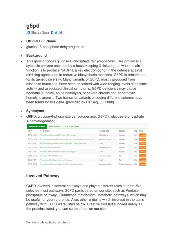
Glucose-6-Phosphate Dehydrogenase (G6PD) deficiency • is the most common human enzyme deficiency in the world; it affects an estimated 400 million people. G6PD deficiency is also known as "favism," since G6PD deficient individuals are also sometimes allergic to fava beans. G6PD deficiency is an allelic abnormality which is inherited in an X-linked recessive fashion
When someone has G6PD deficiency, complications can arise; hemolytic anemia and prolonged neonatal jaundice are the two major pathologies associated with G6PD deficiency. Both of these conditions are directly related to the inability of specific cell types to regenerate reduced nicotinamide adenine dinucleotide phosphate (NADPH); this reaction is normally catalyzed by the G6PD enzyme.
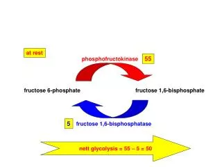
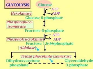
Principle • Glucose-6-phosphate dehydrogenase (G6PDH, D-glucose-6-phosphate) catalyzas the first step in the pentose phosphate shunt ,oxidising glucose-6-phosphate (G-6-P)to 6-phosphogluconate(6-PG) and reducing NADP to NADPH. • G-6-P + NADP+ 6-PG + NADPH + H+ G-6PDH
NADP is reduced by G-6-PDH in the presence of G-6-P. The rate of formation of NADPH is directly proportional to the G-6-PDH activity and is measured spectrophotometrically as an increased in absorbance at 340nm. Prodution of asecond molar equivalant of NADPH by erythrocyte 6-phosphogluconate dehydrogenase (6-PGDH) according to the reaction : • 6-PG + NADP+ Ribulose-5- phosphate + NADPH + H+ + CO2
Specimen collection and storage • Whole blood collected with EDTA, heparine or acid citrate dextrose .Red cell G-6-PDH is stable in whole blood for one week refrigrated (2-8ºc),but is unsteble in red cell hemolysates.
Procedure • prepare reaction mixture: • Add 0.01ml blood directly to vial containing G-6-PDH assay solution and mix throughly to completely suspend erythrocytes, lat stand at room tempreture(18-25ºc) for 5-10min. • Add 2.0ml G-6-PDH substrate solution directly to vial and mix gently by inverting several times. • Transfer contents of vial to cuvet.
Place cuvet in constant tempreture cuvet compartment or water bath and incubate for approximatly 5min to attain therma; equilibrium. • Read and record absorbance (A1) of test at 340 nm vs water or ptassium dichromate solution. This is initial A .(if using awater bath or incubator ,return cuvet to it) • Exactly 5min later, again read and record (A2), this is final A. • To determine G-6-PDH activity do the following calculation.
Calculation • = ΔA per min X 4839 / Hb (g/dl) X TCF Where: • 100 = factor to convert activity to 100ml • 3.01 = total reaction volume (ml) • 0.01 = sample volume (ml) • 6.22 = mill molar absorptive of NADPH at 340 nm • Hb (g/dl) = hemoglobin concentration determined for each specimen • TCF = temperature correction factor (1 at 30ºc)
Qualitative method in G-6-DP determination: • Glucose -6-phosphate dehydrogenase,present in the red blood cell hemoysate, act on glucose -6-phosphate and reduces NADP to NADPH which, with the help of PMS, reduces blue colored 2,6Dichlorophenol Indophenol into acolorless form.the rate of decolorization is proportional to the enzynme activity. The reaction can be represented as: • G-6-phosphate +NADP 6-phosphogluconic acid +NADPH
Procedure: Step1: Preparation of red cell hemolysate: • Purified water : 2.5ml • Fresh blood : 0.05ml • Mix well and allow standing for 5min at R.T.
Step2: Assay of the enzyme: • Add 1mi of the hemolysate (step 1) to the vial of solution 1 and mix gently. • Add immediately about 1ml of reagent 3. • Seal the vial with aluminium foil and incubate in water bath at 37ºc. • Observe: thetime taken for the color change from initial deep blue to reddish purple. Follow up to amax. Of 6 hours with 30 min intervals.
Results Normal: 30-60 min. • G-6-PD deficient (heterozygous males, homozygous female): 140min-24hr • G-6-Pdcarriers (heterozygous females): 90min-several hours.