Corynebacterium
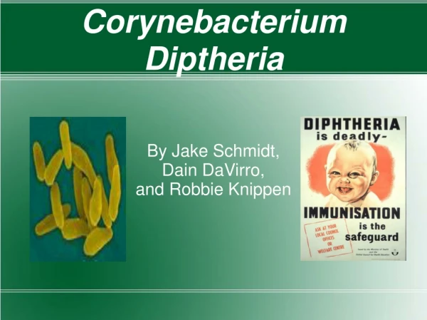
Corynebacterium. Corynebacterium. Classification Corynebacterium diphtheriae and diphtheroids (look like C. diphtheriae ) are Gram- positive, club shaped rods. Some are saprophytic Some produce disease in animals.

Corynebacterium
E N D
Presentation Transcript
Corynebacterium • Classification • Corynebacterium diphtheriae and diphtheroids (look like C. diphtheriae) are Gram- positive, club shaped rods. • Some are saprophytic • Some produce disease in animals. • C. diphtheriae is the most important pathogen in the group. • Morphology and cultural characteristics • Small G+B; arrangement=palisade or Chinese • letters • Growth on B.A – raised, translucent, gray colonies
Corynebacterium • Loeffler‘s agar slant contains serum and egg that enhance the formation of metachromatic granules (polymerizedpolyphosphoric acid) in C. diphtheriae. • Also called Babes-Ernst granules. • They are visualized by staining with methylene blue.
Corynebacterium • A medium containing tellurite should be used to select for Corynebacterium and other G+ organisms -it inhibits G organisms. Two kinds are used: • Cystine tellurite has alonger shelf life • Tinsdale helps to differentiate amongst the Corynebacterium. • Colonies on either appear black or gray due to tellurite reduction. • S. aureus and Listeria also grow as black colonies. • On Tinsdale C. diphtheriae, ulcerans, and pseudotuberculosis form brown halos around the colonies due to formation of ferric sulfide.
Corynebacterium • 3 morphological types of C. diphtheriae are found on tellurite containing media: • Mitis – black colonies with a gray periphery • Gravis – large, gray colonies • Intermedius – small, dull gray to black. • All produce an immunologically identical toxin. • Incubation -35-370 C for 24 hours. • They prefer a pH of 7.8-8.0 for good growth. • They require access to oxygen (poor AnO2 growth). • Biochemistry • Catalase +
Corynebacterium • Nonmotile • C. ulcerans is urease +, C. diphtheriae is -, and C. pseudotuberculosis is usually + • Virulence factors C. diphtheriae • For C. diphtherias to cause diphtheria an exotoxin must be produced. • Is a heat-labile polypeptide produced during lysogeny of a phage that carries the "tox” gene. • Alkaline pH of 7.8- 8.0, aerobic conditions, and a low environmental iron level are essential for toxin production (occurs late in the growth of the organism). • The toxin inhibits protein synthesis by ADP-ribosylating elongation factor 2. • What other organism produces a similar toxin?
Corynebacterium • Trypsin cleaves the toxin into 2 fragments, A and B, that are linked together by a disulfide bridge. • Fragment B is required for toxin binding to tissue cells and fragment A contains the toxic activity. • One molecule of toxin can inhibit 90% of the protein synthesis in a cell. • Systemic effects include heart failure, paralysis and adrenal hypofunction leading to an Addison’s like disease. • C. ulcerans and C. pseudotuberculosis sometimes make a diphtheria-like toxin.
C. diphtheria toxin • Toxin enters through receptor mediated endocytosis • Acidification of endocytic vesicle allows A to dissociate from B • A enters cycoplasm
Corynebacterium • To prove that an isolate can cause diphtheria, one must demonstrate toxin production. • This is most often done on an Elek plate: • The organism is streaked on a plate containing low iron. • A filter strip containing anti-toxin antibody is placed perpendicular to the streak of the organism. • Diffusion of the antibody into the medium and secretion of the toxin into the medium occur. • At the zone of equivalence, a precipitate will form.
Corynebacterium • Guinea pig or tissue culture toxicity assays may also be done. • Capsule – is protein in nature • Cord factor – is a complex glycolipid (trehalose 6,6’-dicorynemycolate) that has been shown to disrupt mouse mitochondria. • It has not been shown to play a role in the production of diphtheria.
Corynebacterium • Clinical Significance (C. diphtheria) • Is normally found in the throats of healthy carriers. • The organism infects only man and it has a limited capacity to invade. • Diphtheria - Disease usually starts as a local infection of the mucous membranes causing a membranous pharyngitis • Local toxin effects result in degeneration of epithelial cells. • Inflammation, edema, and production of a pseudomembrane composed of fibrin clots, leukocytes, and dead epithelial cells and microorganisms occurs in the throat.
Diphtheria - pseudomembrane • This may obstruct the airway and result in suffocation.
Corynebacterium • The more dangerous effects occur when the toxin becomes systemic and attacks the heart(heart failure), peripheral nerves (paralysis), and the adrenal glands (hypofunction). • Cutaneous diphtheria More common in tropical and subtropical areas. • Necrotic lesions with occasional formation of a local pseudomembrane occur. • Antibiotic susceptibility and treatment • Antiserum once the toxin has bound, however, the antiserum against it is ineffective. • Penicillin to eliminate the organism.
Corynebacterium • Prevention- Active immunization with toxoid (alum precipitate). • Is part of the DPT vaccine. • Shick skin test like the Dick test in that it tests for circulating antibody to the toxin by injecting a small amount of toxin intradermally and observing for a local erythematous and necrotic reaction. • If this occurs it indicates that the person has no anti-toxin antibodies and is, therefore, susceptible to diphtheria. • Other Corynebacterium are part of the normal flora of the skin and URT.
Corynebacterium • Are called diphtheroids and may occasionally cause disease, particularly in immunocompromised individuals. • C. ulcerans toxigenic strains may produce a disease similar to, but less severe than diphtheria. • J-KGroup commonly cause infections in those with underlying disease. • Diseases include bacteremia, meningitis, peritonitis, wound infections, etc. • It is becoming more and more of a problem. • C. pseudotuberculosis found in those with exposure to animals. • Can cause pneumonia or lymphadenitis. • Produces a different exotoxin than C. diphtheriae.