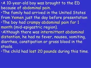
A 10-year-old boy was brought to the ED because of abdominal pain. The family had arrived in the United States from Ye
A 10-year-old boy was brought to the ED because of abdominal pain. The family had arrived in the United States from Yemen just the day before presentation The boy had crampy abdominal pain for 1 month (mid-epigastric region).

A 10-year-old boy was brought to the ED because of abdominal pain. The family had arrived in the United States from Ye
E N D
Presentation Transcript
A 10-year-old boy was brought to the ED because of abdominal pain. • The family had arrived in the United States from Yemen just the day before presentation • The boy had crampy abdominal pain for 1 month (mid-epigastric region). • Although there was intermittent abdominal distention, he had no fever, nausea, vomiting, diarrhea, constipation or gross blood in the stools. • The child had lost 20pounds during this time.
PMH: • Received immunizations, but no documentation was available • No hospitalization or surgery • History of contact with cows and goats and a questionable consumption of unpasteurized milk. • The water supply was reportedly clean • No history of swimming in fresh water. • The patient walked barefoot outside. • No household members had similar symptoms.
Physical examination • A thin, cooperative boy in no acute distress. V/S were NL. • H&N: normal. No lymphadenopathy • Heart: regular rate and rhythm, with no murmurs. • The lungs were clear to auscultation bilaterally. • The abdomen was soft, but mildly distended, and bowel sounds were present. There was no rebound tenderness, guarding, ascites or masses. The liver was nontender and smooth, with the edge palpable 8 cm below the right costal margin. The spleen was not palpable. • The rest of the physical examination was unremarkable.
Labs • WBC: 15 000/mm3 (4% bands, 8%N, 41% L, 2% M, 44% E and 1% B). • HB 12.9 g/dl, platelet count was 494K • Serum electrolytes, coagulation profile, liver enzymes, bilirubin and urine analysis were normal. • Computed tomography of the abdomen showed hepatomegaly. • Esophagogastroduodenoscopy revealed esophagitis, but no varices.
Hepatitis A and hepatitis B serology indicated past infection. • Hepatitis C IgG : negative. • Serum immunoglobulins: • Normal IgM, • Slightly elevated IgA and IgG. • IgE was 23 010 IU/ml, 10 times the upper end of normal (normal 1 to 240 IU/ml).
Serology for Toxocaraspecies and Trichinella spiralis was negative. • Filariasis was excluded. • There were no ova or parasites in the stools on three separate occasions. • Stool and urine examinations were negative for schistosomiasis. • The diagnosis was made only after a more invasive study.
Because this boy had • Persistent gastrointestinal complaints • Came from an endemic area • Walked barefoot • Had eosinophilia • Extremely elevated serum IgE value, schistosomiasis was suspected and a rectal biopsy was done.
The biopsy showed ellipsoid eggs, each with a lateral spine, characteristic of Schistosoma mansoni .Pediatr Infect Dis J. 1999 Jun;18(6):556, 572-3
Schistosomiasis Nahed Abdel-Haq, M.D Division of Infectious Diseases Children’s Hospital of Michigan
Editorial Mind over matter: Heroes overcome special needs Chris Burke A hero with trisomy-21. A question was raised by many of The Ambassadors Magazine's readers. Why not dedicate an issue of the MEGASTARS section to people with special needs? In previous issues we've focused on and reported several stories about individuals who have succeeded over debilitating ailments to accomplish, in some cases, remarkable achievements. One such story was that of Goodwill Ambassador for the National Down Syndrome Society, Chris Burke. Other notables with trisomy-21 whose lives were explored in the pages of the Ambassadors include Nigel Hunt (England), Sheenagah Hardie (Scotland) and Omar A. Al-Awadi (Kuwait).This issue's feature story is no different. This time we look at 1.5 million Americans who are affected by short stature and some of their achievements in all facets of life despite all obstacles. This section, however, is reserved for a tribute to a selection of persons across time and place who have defeated their 'special needs' and proved that resilience and ambition can help conquer most obstacles. President Franklin D. Roosevelt defeating poliomyelitis President FDR with Eddie Cantor President Franklin D. Roosevelt (FDR), himself a polio survivor, was confined to a wheelchair most of his later years. However, this obstacle didn't stop him from pursuing and achieving the highest seat in the nation. A much-loved, politician, strategic tactician, able diplomat and devoted leader, FDR dedicated much of his efforts to supporting polio research, raising in 1937 $1-million at the annual Presidential ball alone. Roosevelt's friend, celebrity Eddie Cantor, recommended that he continue the fund-raising by asking each American to contribute a dime "The March of Dimes." And so, in one of FDR's famous weekly radio addresses, he asked each citizen to send a dime to the White House to help fund polio-research. 1938, Roosevelt created the National Foundation of Infantile Paralysis (March of Dimes), was the name used for the fund-raising itself and the foundation mission was to prevent and cure polio. The foundation supported the discovery of both Salk & Sabin's vaccines for polio prophylaxis. The March of Dimes foundation continued its medical research towards other disabling conditions, as a result of which, ten of its scientists have become Noble Prize laureates. President FDR, who was stricken with the polio virus when he was 39, affecting both his lower limbs causing flaccid paraplegia, succeeded to regain the use of his legs through swimming. As a direct result of FDR's initiative, polio is now vanishing all over the world. Eng and Cheng: The Famous Siamese Twins (1811-1874) Few birth defects have become as sensationalized more than Siamese twins, and none more popularized than the astonishing story of Eng and Cheng. On May 11, 1811 Chang and Eng were born in China, where there was much excitement among the midwives and neighbors attending the historic birth when they learned that the twins were connected by a short, flexible, fleshy band or ligament, breastbone to breastbone. It wasn't long before the initial excitement turned to fright and no one would touch them. However, their mother appears to have been quite sensible. She untwisted them and straightened the band, and bathed them. Thus began the life of the famous conjoined twins who gave the world the term "Siamese twins". The consensus among the doctors was that the twins could not be separated.. Sallie, Eng, Cheng, Adelaide, and sons Patrick Henry and Albert (1865). Chang and Eng moved to USA and soon became good friends with a neighbor, and eventually married two of his daughters (Sallie and Adelaide). Originally they setup housekeeping in one house, but as the children started coming, one household was not big enough. Chang and Adelaide had ten children. Eng and Sallie had eleven children. Eventually they moved into separate houses spending three days at each other's house. In January 1874, Chang died during the night. The next morning, Sallie sent one of the children racing to get Dr. Hollingsworth to separate Eng from Chang. A formerly healthy Eng complained of being "very bad off." He was rational but terrified and complained of agonizing pain and distress, especially in his limbs. Finally, he lapsed into a coma-like stupor. An hour later, he died at 63 years of age. The uniqueness of End and Cheng's story and their ability to forge somewhat normal lives is in itself a remarkable achievements. The Blind Mountain-climber Erik Weihenmayer Napoleon after coronation as Emperor of France (Painting by Ingres) Abdel-Halim Hafez, the popular Arabic musician and singer Prof. Talaat I. Farag, MD, FRCP(Edin), FACPChief Editor, The Ambassadors MagazineTel/Fax: (902) 425-0375Email: mail@ambassadors.net Erik Weihenmayer (left) Abdel-Halim Hafez, the popular Arabic musician and singer Napoleon after coronation as Emperor of France What could they have in common? Akhnaton, was allegedly diagnosed with bilharziasis or schistosomiasis. Some also mentioned that Napoleon Bonaparte was infected with this parasite after swimming in Egypt. The famous Egyptian singer, Abdel Halim Hafez died with complications of this disease On June 18, 2001, TIME magazine chose 33-year-old to be the subject of a special feature story and put the blind hero on its issue cover. For some persons just crossing the street can be a risky venture. Erik Weihenmayer, who was the victim of a rare hereditary disease of the retina (retinoschis), causing his blindness since he was thirteen. This year Erik became the first sightless person to scale Mount Everest's killer peak, a heroic accomplishment by every measure. Plenty of sighted people walk through life with less poise and grace than Erik, unsure of their steps, and second-guessing their every move. On his Everest climb, Erik humbly exclaims that "If a blind guy can do it, anyone can." But even the many able-sighted weren't as lucky as Erik. Almost 90% of Everest climbers have failed to reach the summit. Horrifyingly, since 1953, at least 165 never returned from the climb. Erik and his wife Ellie and their one-year-old daughter, Emma, celebrated his climb. Akhnaton, Napoleon, and Abdel Halim Hafez An ancient Egyptian pharaoh, a French emperor and an Arabic musical icon. What could they have in common? The Ancient Egyptian Pharaoh, Akhnaton, was allegedly diagnosed with "AAA-disease" (known as bilharziasis or schistosomiasis). This endemic parasitic disease was first described in Cairo by the German scientist, Theodor Bilharz in 1851. Some also mentioned that Napoleon Bonaparte was infected with this parasite after swimming in Egypt. It is well known that the famous Egyptian singer, Abdel Halim Hafez died with complications of this disease. Bilharziasis has high prevalence among rural farmers and can cause liver cirrhosis, enlarged spleen, cancer bladder, etc. With the disease's victims dating back 4,000 years, the Egyptian government launched a mass campaign headed by Prof. Ismail Sallam (Minister of Health and Population) to eradicate the disease. During my recent visit to Egypt to offer lectures to family doctors in four provinces, I found that the prevalence of bilharziasis in Munofia province has dropped to less than 2%. Our sincere congratulations and acknowledgement to all those who have contributed to this campaign. By presenting the brief stories of persons with different special needs, from Akhnaton to Roosevelt it is clear that a dream can conquer any and all involuntary limitations. We have to search together in order to find preventive tools for all environmental and genetic problems which could affect our children and grandchildren while continuing to encourage those around us who suffer with such ailments to aspire and dream. Below, I have borrowed from the words of the acknowledged poet Hala El-Banna to inspire us all to search the depths for the pearls within: I'll keep searching in the deepest ocean,I'll keep looking everywhere I go,I'll keep hoping for that ray of light;the one that'll brighten my days and nights.I'll keep dreaming, for maybe one dayI'll reach a long sought dreamAnd turn abstract into concrete The Ambassadors
Schistosomiasis • Described by Theodore Bilharz in Cairo in 1851 (Bilharzia) • 200 M people in 74 countries • 120 M have symptoms • 20 M have severe illness • Despite control programs: continues to spread • Reports of R to praziquantil Allen GP et al. NEJM 2002, Volume 346:1212-1220
Schistosomiasis is second only to malaria in human impact among tropical diseases and is the third most prevalent parasitic disease in the world.
Schistosomiasis/Etiology • Human disease: caused mainly by 3 species of flat worms • S. mansoni: most common (Africa), intestinal disease • S. japonicum: Asia, Pacific, intestinal disease • S. haematobium: affects 54 countries in Africa and the Middle East, urinary disease
Schistosomiasis/Unusual species • S. mekongi: disease mainly in the Mekong river basin, • Related to S. japonicum • Disease is similar, milder • S. intercalatum: in central Africa • Similar to S. manosoni but milder • S. mattheei and S. bovis: mainly animals
Schistosomiasis/Distribution • Schistosoma haematobium, S. mansoni and S. intercalatuminfections: in sub-Saharan Africa • S. mansoni remains endemic in parts of Brazil, Venezuela and the Caribbean • S. japonicum still occurs in China, Indonesia & the Philippines • S. makongi: Cambodia & Laos (Mekong River)
Schistosomiasis/Epidemiology • Humans are principal hosts for major species • Intermediate host is the snail • Appropriate snail is required for maintaining the cycle • Eggs excreted in stools: S. mansoni, S. japonicum • Eggs excreted in urine: S. haematobium • Eggs hatch in fresh water into motile miracidia which infect snails
Life Cycle of the Schistosome Allen GP et al. NEJM 2002, Volume 346:1212-1220
Adult worms: males/females Cercariae: infectious stage
In endemic areas, most at risk are school-age children, women, and those involved in occupations such as irrigation, farming and fishing.
Epidemiology/travelers • Most in travelers to Africa • Swimming, wading, bathing in fresh water in areas of poor sanitation, snail hosts • Most cases are in swimmers in • Lake Malawi • Lake Kariba • Zambezi River • Present with acute schsitosomiasis • Common early symptoms: hematurea, diarrhea • Rare: transverse myelitis
Epidemiology/Immigrants • Immigrants from endemic areas: may remain infected for 30-40 yrs • Average life span of schistosome is 5 yrs • Adult worms may live for decades • Not notifiable disease in US • True incidence in immigrants, returned travelers is unknown
Epidemiology/Animals • Various animals, such as dogs, cats, rodents, pigs, horses and goats, serve as reservoirs for S. japonicum, and • Dogs for S. mekongi • Variants in birds
Susceptibility • HLA class I and II antigens : more severe manifestations of the disease • HLA-B16 and Cw2: S. haematobium associated bladder CA in Egypt • HLA-DR, DQ, DP: protection from hepatic fibrosis • Advanced fibrosis is related to gamma-interferon receptor gene on chromosome 6 • Resistance to re-infection: 5q31-q35
Schistosomiasis/clinical manifestations • Maculopapular eruption at site of penetration of cercariae • Develops few hours after infection • May develop up to one week later • Similar but less severe than swimmer’s itch
Schistosomiasis/clinical manifestations • Swimmer’s itch: sensitized individuals exposed to non-human schistosomes • Various avian and mammalian schistosomes cause the reaction • Mild-moderate pruritus at penetration site in few hours • Intermittent pruritus, papular eruption in 5-14 d • The cycle of infection is not completed in humans: no systemic complications
Acute Schistosomiasis/Katayama Fever • Areas with high transmission rates • Immune complex-mediated reaction • Deposition of eggs in tissues • Contaminated water exposure 14-84 days earlier • All patients have eosinophilia • Not all shed eggs
Acute Schistosomiasis/Katayama Fever • Fever, headache, generalized myalgias • Right upper quadrant pain, tender hepatomegaly • Bloody diarrhea • Respiratory symptoms: 70% S. mansoni • Interstitial pneumonia (radiologic) • Splenomegaly: 30% • Aseptic meningitis
Chronic Schistosomiasis • A results of host immune response to schistosome eggs • Granulomatous reaction to secreted Ag/fibrosis • Severity depends on: • Amount of Ag release (severity & duration) • Intensity of fibro-obstructive disease • Granulomas at sites of maximum egg deposition • S. mansoni & S. japonicum: intestine & liver • S. haematobium: genitourinary tract
Chronic Schistosomiasis • Granulomas may develop in other organs: • Skin • Lungs • Brain • Adrenal glands • Skeletal muscles • The inflammatory response assist in migration of the eggs to the lumen (GI,UB) • Egg output is low in immundeficient pts
Chronic SchistosomiasisGI • Gut wall: inflammation, hyperplasia, ulcers, microabscesses, polyposis • Colicky pain: lower abdomen, left iliac fossa • Diarrhea: common, + constipation • Blood: occult, gross • Colonic, rectal stenosis • Colonic polyposis:protein-losing enteropathy • Colorectal CA risk: small; if any
Chronic SchistosomiasisLiver • Embolization of eggs to the liver • S. mansoni and S. japonicum • Presiusoidal inflammation, periportal fibrosis • Incidence: 4-8% of pts • Takes yrs, heavy infection • Hepatomegaly: granulomatous inflamm., early • Periportal collagen: • Obstruct blood flow • Portal hypertension • Varices, bleeding, splenomegaly
Chronic SchistosomiasisLiver • Periportal fibrosis seen by US, CT, MRI • Liver synthetic function is preserved until late • Lobular architecture is retained • No nodular regenerative hyperplasia • Quantification of hepatosplenic disease: clinical, US criteria by WHO • S. haematobium: occasional colonic, hepatic disease
Chronic SchistosomiasisLiver • Coinfection with HBV, HCV may occur • S. mansoni: accelerated deterioration • Higher risk of hepatocellular CA (HBV) • S. japonicum: no significant interaction • EGYPT: • Mass campaign of parenteral therapy for schstosomiasis • High prevalnce of HCV in the country: widespread reuse of needles
The abdomen of an 11-year-old boy with intestinal schistosomiasis with the size and extent of the liver and spleen marked, indicating the severity of infection. The disease has caused a stunting of the boy's growth, he is only 120cms tall and weighs 22 kg. WHO
Lancet Infect Dis. 2004 Aug;4(8):498. Clinical picture Schistosomal appendicitis Eric Adehossi and Philippe Parola , Service des Maladies Infectieuses et Tropicales, CHU Nord, Marseille, France A 24-year-old African man recently emigrated to France from Sierra Leone was admitted to the department of surgery of our hospital for a 2-day history of acute abdominal pain. Physical examination of the abdomen revealed right iliac fossa tenderness and guarding, and peritonism. Standard blood haematology and chemistry values were within normal limits, except an increased C-reactive protein (30mg/L). Increased abdominal pain without any fever was noticed during the first 2 days of hospitalisation. Abdominal computed tomography scan showed calcifications from the initial to the distal part of both ureters, in the appendix (figure A), in the seminal vesicles, and within the ureteral wall of the bladder. This presentation is typical of genitourinary schistosomiasis due to Schistosoma haematobium, which is endemic in Sierra Leone, with schistosomial appendicitis. Neither eggs nor other parasites were detected in the urine and stools.
Abdominal CT scan showed calcifications from the initial to the distal part of both ureters and in the appendix Histological examination of the appendix showed numerous S haematobium eggs in submucosa of the appendix
Chronic SchistosomiasisGenitourinary • Specific for S. haematobium • Hematurea: first sign (10-12wks after infection) • Graulomatous response: eggs in tissue • Dysurea: early and late in disease
GenitourinaryLate manifestations • Proteinurea (nephrotic) • Bladder calcification • Obstruction of ureter • Renal colic • Hydronephrosis • Renal failure • secondary bacterial infection • Cystoscopy: areas of rough mucosa (eggs) • Structural urinary tract abnormalities
GU Schstosomiasis. Plain radiograph showing: Calcification of the distal two thirds of both ureters and Bladder calcifications.(The Encyclopaedia of Medical Imaging Volume VII.)
Bladder Schstosomiasis. IVP showing Filling defects in the urinary bladder secondary to granulomas. (The Encyclopaedia of Medical Imaging Vol.VII.)
Genital schistosomiasis • 1/3 of infected women • Isolated internal genital disease is rare • Vulva, perinium: ulcers, fistulas, hypertrophic, wart-like lesions • Tubular infertility: rare, late • May facilitate transmission of HIV
Schistosomiasis/Neurologic • Egg deposits in the CNS: aberrant migration of worms, embolization • Not all are symptomatic • Focal or generalized seizures are typical for S. japonicum CNS involvement • Focal neurologic deficits • 4.3% of hospitalized Chinese adults have CNS disease
Schistosomiasis/Neurologic • Transverse myelitis: most common neurologic complication of S. mansoni and S. haematobium Treatment • Supportive • Steroids • Anticonvulsants: long term use rarely needed
Schistosomiasis • In childhood: growth retardation, anemia • Cognitive impairment, memory deficits • May affect maternal and fetal health • Praziquantel: category B (safe in animals, not tested in humans) • Risk:benefit: treat esp. after 4th month of gestation
Two boys, victims of schistosomiasis showing typical distension of the abdomen. WHO
GenitourinaryLate manifestations • S. haematobium has role in some bladder CA • Squamous cell carcinoma (SCC) • In Egypt: SCC 18-28% of all CA • Mainly male, smokers • The finding is not consistent in all countries with endemic S. haematobium
Schistosomiasis/Differential diagnosis • Toxocara infection • Trichinella spiralis infection • Filariasis • Hepatitis B, C virus infection • Tuberculosis • Amebiasis • Leishmaniasis • Myeloproliferative disease • Peptic ulcer disease • Cancer (GI, GU)