Download
1 / 21
210 likes | 233 Vues
Explore how proteins are regulated through proteolytic activation, with examples in digestive enzymes, blood clotting, and other biological processes like apoptosis and collagen breakdown.

E N D
Lecture 17:Regulation of Proteins 4:Proteolytic Activation Examples Activation of Digestive Enzymes Blood Clotting
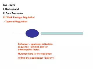
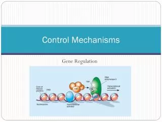
Biological Processes are Carefully Regulated Allosteric Control: The activity of some proteins can be controlled by modulating the levels of small signalling molecules. The binding of these molecules causes conformational changes in the protein which affect its activity. Multiple forms of Enzymes: Different tissues or developmental stages sometimes have specific versions of a given enzyme which have distinct properties although they may have the same basic activity. Reversible Covalent Modification: The activity of many proteins is controlled by attachment of small chemical groups. The most common such modification is phosphorylation- attachment of a phosphate group. Proteolytic Activation: Some enzymes are synthesized in an inactive form and must be activated by cleavage of the inactive form.
Zymogens Some enzymes are synthesized in an initially inactive (but folded) form which is converted to an active form by specific proteolytic cleavage. These initial forms are called zymogens or proenzymes. This method of regulation does not require an energy source unlike phosphorylation which requires ATP. Therefore extracellular enzymes may be activated by this process. Proteolysis is irreversible- once activated, the molecule remains in the activated state.
Examples of Proteolytic Activation Digestive Enzymes: The primary enzymes that function in breaking down proteins and peptides during digestion are synthesized as zymogens in the stomach and pancreas. Blood Clotting: Rapid response to injury is possible by activating a cascade of zymogens.
Hormones: Some hormones, e.g. insulin, are synthesized as precursors which must be activated by proteolysis. Collagen: The major component of skin and bone, collagen is derived from its precursor procollagen by specific proteolysis. Developmental Processes: The structural protein collagen must be broken down in certain tissues at particular stages during normal development. The protease responsible for this process, collagenase, is activated at the precise time needed by specific proteolysis. Apoptosis: Cells have an intrinsic ability to “self-destruct.” This process, programmed cell death or apoptosis, is required during normal development and also functions to eliminate cells that are somehow damaged, eg infected with pathogens or containing DNA too damaged to repair. This process is mediated by proteolytic enzymes called caspases, which are initially synthesized as inactive procaspases and can be activated by proteolysis in response to a variety of signals.
Digestive Zymogens The pancreas is a major producer of digestive enzymes. Acinar cells in the pancreas produce a variety of zymogens which are stored in membrane-bounded granules. These zymogen granules fuse with the cell membrane in response to signals from hormones or nerve impulses, releasing their contents into ducts leading to the digestive tract. The zymogens include trypsinogen, chymotrypsinogen, proelastase, and procarboxypeptidase.
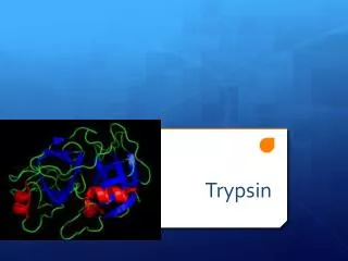
Activation of Digestive Zymogens The different digestive proteases have different substrate specificities, enabling the breakdown of a wide variety of peptides. The zymogens are all activated by a single enzyme, trypsin. Trypsin itself is activated by enteropeptidase, which is secreted by cells lining the digestive tract. In turn trypsin activates the other zymogens.
Activation of Chymotrypsin Chymotrypsin is initially synthesized as the inactive precursor chymotrypsinogen. Initial cleavage by trypsin yields p-chymotrypsin, which is further processed by chymotrypsin itself to yield a-chymotrypsin, the final active form.
Structural Basis of Chymotrypsin Activation Comparison of the structures of chymotrypsin and chymotrypsinogen revealed that the inactive and active forms are very similar overall but that small, local rearrangements exist that explain the difference in activity. The break at Ile 16 creates a new positive amino terminus which forms a buried ionic interaction with Asp 194. Subsequent rearrangements cause the formation of a hydrophobic cavity important for substrate specificity, and also formation of the oxyanion hole which is required for the the catalytic activity of the activated enzyme.
Inhibition of Trypsin The accidental activation of a few trypsin molecules inside the acinar cells could be disastrous. A small amount of active trypsin could activate all the zymogens which would lead to digestion of all the proteins in the cell. To guard against this possibility, the acinar cells contains a small (6 kD )protein that inhibits trypsin- pancreatic trypsin inhibitor or PTI. PTI binds extremely tightly to trypsin- even 8M urea or 6M HCl cannot dissociate the complex. The tight binding is partly conferred by a Lys side-chain which binds in a negatively charged pocket on trypsin. PTI is eventually cleaved by trypsin but only extremely slowly (over months) and the combination of tight binding and slow hydrolysis makes it a very effective inhibitor.
Emphysema Emphysema can result from a defect in a similar type of inhibitor. Emphysema is a result of loss of elasticity in the alveolar walls of the lungs, reducing the volume in the lungs available for exchange of O2 and CO2. This loss of elasticity is caused by damage to elastic fibers, composed of connective tissue proteins. White blood cells secrete elastase, which is a protease that is capable of degrading elastic fibers. Normally this is prevented by a protein in blood plasma called a1-antiproteinase that binds to and inhibits the secreted elastase, protecting your lungs from damage. People with inherited disorders in this inhibitor or its production (it is secreted by the liver) are at much higher risk for developing emphysema. There is a family of such inhibitors, called serpins, which is short for Serine Protease Inhibitors.
Connection between Smoking and Emphysema Tobacco smoke contributes to emphysema by damaging a1-antiproteinase- the smoke oxidizes a particular methionine residue on a1-antiproteinase: Methionine sulfoxide Methionine This residue is an essential part of the recognition interface between elastase allowing it to bind a1-antiproteinase. When this methionine is oxidized, the binding is disrupted, the a1-antiproteinase can no longer inhibit elastase, and elastase degrades the elastic fibers in the lungs, leading to emphysema. Smoking is particularly dangerous for persons with a genetic defect in the inhibitor.
Activation Cascades Rapid response to a stimulus is possible through a cascade of enzyme activations. A cascade consists of a series of several steps each of which has a multiplicative effect on subsequent steps. Step 1: A signalling molecule activates 1 molecule of enzyme 1. Step 2: Enzyme 1 activates 100 molecules of enzyme 2. (100-fold amplification) Step 3: Each activated molecule of enzyme 2 activates 100 molecules of enzyme 3. (104-fold activation) Step 4: Each activated molecule of enzyme 3 activates 100 molecules of enzyme 4. (106-fold activation) Cascades can produce an enormous and extremely rapid response. An example of such a process occurs in blood clotting.
Blood Clotting: A Cascade of Zymogen Activations The clotting of blood after injury must be rapid to avoid blood loss. The rapidity with which this is accomplished is due to a cascade of activation of blood clotting factors. Small amounts of the initial clotting factors amplify the response and result in the rapid formation of clots. Clotting factors are referred to by Roman numerals. These were named in the order that they were discovered, not for the order in which they act. The inactive zymogen form is denoted by the Roman numeral, (e.g. Factor X) and the activated form is indicated by adding the suffix “a”. (e.g. Factor Xa)
Two Pathways of Blood Clotting The blood-clotting cascade can be activated in two different ways. The intrinsic pathway is initiated by exposure of abnormal surfaces of ruptured blood vessels. The extrinsic pathway is initiated by trauma, resulting in the by the release of Tissue factor, a lipoprotein. Both pathways converge in the final steps, in which the protease thrombin is activated and releases the clot-forming protein fibrin from its precursor fibrinogen. Hemophilia results from the loss of Factor VIIIa, which partially or wholly blocks the intrinsic pathway. The resulting inability to form clots can make even a small wound life-threatening.
Final Steps in Clot Formation Clots consist largely of ordered fibrous arrays of the protein fibrin. Fibrin is cleaved from its zymogen fibrinogen by the protease thrombin. When released from fibrinogen, fibrin rapidly polymerizes into ordered arrays. These arrays are further stabilized by covalent crosslinks between fibrin monomers. Activation Fibrin release Crosslinking
Fibrinogen and Fibrin Fibrinogen constitutes 2-3% of blood plasma protein. It exists as a complex of 3 subunits Aa, Bb, and g. Small peptides A and B are removed by thrombin to release fibrin, revealing creating new termini which enable fibrin to polymerize into fibers. Fibrinogen
Clot Formation by Fibrin The new termini of the a chain created when the A peptides are cleaved off by thrombin interact with binding sites on the g subunit. The fibers are further stabilized by amide crosslinks between fibrin monomer side-chains. Binding site Fibrin array and electron micrograph g Transglutaminase
Cessation of Clot Formation The cascade of activations during clot formation must be carefully regulated so that clots will not continue to expand more than necessary, which would block blood flow to healthy tissue (thrombosis). Once initiated, the clotting cascade is attenuated by loss of clotting factors through dilution, removal from the bloodstream, and by proteolysis. Specific inhibitors to individual clotting factors (serpins) exist which also attenuate the cascade. Protein C is a protease that degrades factors Va and VIIIa. It is activated by thrombin. Once the final steps of the cascade are reached, the factors carrying out the prior steps are deactivated.
Removal of Clots When no longer required clots are removed by proteolysis of fibrin by the protease plasmin. Plasmin is itself originally produced as an inactive precursor, plasminogen, which is released through the action of tissue-type plasminogen activator (TPA). TPA is given to some heart attack victims to restore circulation through blocked blood vessels. Blood flow restored: Blockage removed after TPA was administered Blood vessel in heart blocked by clot
Summary: Zymogens are inactive protein precursors which must be converted to their active forms by specific proteolytic cleavage events. A variety of digestive enzymes are synthesized as zymogens in the pancreas. They are activated by proteolysis, and further control of their activities is achieved through the action of specific inhibitor proteins. A cascade of zymogen activations resulting in the controlled creation of fibrin aggregates is the molecular basis of blood clotting. Key Concepts: Zymogens Control of activation Roles of inhibitor proteins (Serpins) Emphysema Activation Cascades Mechanism of blood clotting Hemophilia