Download
1 / 70
940 likes | 2.94k Vues
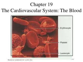
Chapter 19 The Cardiovascular System: The Blood . Fluids of the Body . Cells of the body are serviced by 2 fluids blood composed of plasma and a variety of cells transports nutrients and wastes interstitial fluid bathes the cells of the body

E N D
Chapter 19The Cardiovascular System: The Blood Tortora & Grabowski 9/e 2000 JWS
Fluids of the Body • Cells of the body are serviced by 2 fluids • blood • composed of plasma and a variety of cells • transports nutrients and wastes • interstitial fluid • bathes the cells of the body • Nutrients and oxygen diffuse from the blood into the interstitial fluid & then into the cells • Wastes move in the reverse direction • Hematology is study of blood and blood disorders Tortora & Grabowski 9/e 2000 JWS
Functions of Blood • Transportation • O2, CO2, metabolic wastes, nutrients, heat & hormones • Regulation • helps regulate pH through buffers • helps regulate body temperature • coolant properties of water • vasodilatation of surface vessels dump heat • helps regulate water content of cells by interactions with dissolved ions and proteins • Protection from disease & loss of blood Tortora & Grabowski 9/e 2000 JWS
Physical Characteristics of Blood • Thicker (more viscous) than water and flows more slowly than water • Temperature of 100.4 degrees F • pH 7.4 (7.35-7.45) • 8 % of total body weight • Blood volume • 5 to 6 liters in average male • 4 to 5 liters in average female • hormonal negative feedback systems maintain constant blood volume and osmotic pressure Tortora & Grabowski 9/e 2000 JWS
Techniques of Blood Sampling • Venipuncture • sample taken from vein with hypodermic needle & syringe • median cubital vein (see page 717) • why not stick an artery? • less pressure • closer to the surface • Finger or heel stick • common technique for diabetics to monitor daily blood sugar • method used for infants Tortora & Grabowski 9/e 2000 JWS
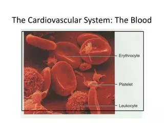
Components of Blood • Hematocrit • 55% plasma • 45% cells • 99% RBCs • < 1% WBCs and platelets Tortora & Grabowski 9/e 2000 JWS
Blood Plasma • 0ver 90% water • 7% plasma proteins • created in liver • confined to bloodstream • albumin • maintain blood osmotic pressure • globulins (immunoglobulins) • antibodies bind to foreignsubstances called antigens • form antigen-antibody complexes • fibrinogen • for clotting • 2% other substances • electrolytes, nutrients, hormones, gases, waste products Tortora & Grabowski 9/e 2000 JWS
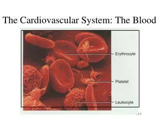
Formed Elements of Blood • Red blood cells ( erythrocytes ) • White blood cells ( leukocytes ) • granular leukocytes • neutrophils • eosinophils • basophils • agranular leukocytes • lymphocytes = T cells, B cells, and natural killer cells • monocytes • Platelets (special cell fragments) Tortora & Grabowski 9/e 2000 JWS
Hematocrit • Percentage of blood occupied by cells • female normal range • 38 - 46% (average of 42%) • male normal range • 40 - 54% (average of 46%) • testosterone • Anemia • not enough RBCs or not enough hemoglobin • Polycythemia • too many RBCs (over 65%) • dehydration, tissue hypoxia, blood doping in athletes Tortora & Grabowski 9/e 2000 JWS
Blood Doping • Injecting previously stored RBC’s before an athletic event • more cells available to deliver oxygen to tissues • Dangerous • increases blood viscosity • forces heart to work harder • Banned by Olympic committee Tortora & Grabowski 9/e 2000 JWS
Formation of Blood Cells • Most blood cells types need to be continually replaced • die within hours, days or weeks • process of blood cells formation is hematopoiesis or hemopoiesis • In the embryo • occurs in yolk sac, liver, spleen, thymus, lymph nodes & red bone marrow • In adult • occurs only in red marrow of flat bones like sternum, ribs, skull & pelvis and ends of long bones Tortora & Grabowski 9/e 2000 JWS
Hematopoiesis Tortora & Grabowski 9/e 2000 JWS
Stages of Blood Cell Formation • Pluripotent stem cells • .1% of red marrow cells • replenish themselves as they differentiate into either myeloid or lymphoid stem cells • Myeloid stem cell line of development continues: • progenitor cells(colony-forming units) no longer can divide and are specialized to form specific cell types • example: CFU-E develops eventually into only red blood cells • next generation is blast cells • have recognizable histological characteristics • develop within several divisions into mature cell types • Lymphoid stem cell line of development • pre-B cells & prothymocytes finish their develop into B & T lymphocytes in the lymphatic tissue after leaving the red marrow Tortora & Grabowski 9/e 2000 JWS
Hemopoietic Growth Factors • Regulate differentiation & proliferation • Erythropoietin (EPO) • produced by the kidneys increase RBC precursors • Thrombopoietin (TPO) • hormone from liver stimulates platelet formation • Cytokines are local hormones of bone marrow • produced by some marrow cells to stimulate proliferation in other marrow cells • colony-stimulating factor (CSF) & interleukin stimulate WBC production Tortora & Grabowski 9/e 2000 JWS
Medical Uses of Growth Factors • Available through recombinant DNA technology • recombinant erythropoietin (EPO) very effective in treating decreased RBC production of end-stage kidney disease • other products given to stimulate WBC formation in cancer patients receiving chemotherapy which kills bone marrow • granulocyte-macrophage colony-stimulating factor • granulocyte colony stimulating factor • thrombopoietin helps prevent platelet depletion during chemotherapy Tortora & Grabowski 9/e 2000 JWS
Red Blood Cells or Erythrocytes • Contain oxygen-carrying protein hemoglobin that gives blood its red color • 1/3 of cell’s weight is hemoglobin • Biconcave disk 8 microns in diameter • increased surface area/volume ratio • flexible shape for narrow passages • no nucleus or other organelles • no cell division or mitochondrial ATP formation • Normal RBC count • male 5.4 million/drop ---- female 4.8 million/drop • new RBCs enter circulation at 2 million/second Tortora & Grabowski 9/e 2000 JWS
Hemoglobin • Globin protein consisting of 4 polypeptide chains • One heme pigment attached to each polypeptide chain • each heme contains an iron ion (Fe+2) that can combine reversibly with one oxygen molecule Tortora & Grabowski 9/e 2000 JWS
Transport of O2, CO2 and Nitric Oxide • Each hemoglobin molecule can carry 4 oxygen molecules from lungs to tissue cells • Hemoglobin transports 23% of total CO2 waste from tissue cells to lungs for release • combines with amino acids in globin portion of Hb • Hemoglobin transports nitric oxide & super nitric oxide helping to regulate BP • iron ions pick up nitric oxide (NO) & super nitric oxide (SNO)& transport it to & from the lungs • NO causing vasoconstriction is released in the lungs • SNO causing vasodilation is picked up in the lungs Tortora & Grabowski 9/e 2000 JWS
RBC Life Cycle • RBCs live only 120 days • wear out from bending to fit through capillaries • no repair possible due to lack of organelles • Worn out cells removed by fixed macrophages in spleen & liver • Breakdown products are recycled Tortora & Grabowski 9/e 2000 JWS
Recycling of Hemoglobin Components • In macrophages of liver or spleen • globin portion broken down into amino acids & recycled • heme portion split into iron (Fe+3) and biliverdin (green pigment) Tortora & Grabowski 9/e 2000 JWS
Fate of Components of Heme • Iron(Fe+3) • transported in blood attached to transferrin protein • stored in liver, muscle or spleen • attached to ferritin or hemosiderin protein • in bone marrow being used for hemoglobin synthesis • Biliverdin (green) converted to bilirubin (yellow) • bilirubin secreted by liver into bile • converted to urobilinogen then stercobilin (brown pigment in feces) by bacteria of large intestine • if reabsorbed from intestines into blood is converted to a yellow pigment, urobilin and excreted in urine Tortora & Grabowski 9/e 2000 JWS
Erythropoiesis: Production of RBCs • Proerythroblast starts to produce hemoglobin • Many steps later, nucleus is ejected & a reticulocyte is formed • orange in color with traces of visible rough ER • Reticulocytes escape from bone marrow into the blood • In 1-2 days, they eject the remaining organelles to become a mature RBC Tortora & Grabowski 9/e 2000 JWS
Feedback Control of RBC Production • Tissue hypoxia (cells not getting enough O2) • high altitude since air has less O2 • anemia • RBC production falls below RBC destruction • circulatory problems • Kidney response to hypoxia • release erythropoietin • speeds up development of proerythroblasts into reticulocytes Tortora & Grabowski 9/e 2000 JWS
Normal Reticulocyte Count • Should be .5 to 1.5% of the circulating RBC’s • Low count in an anemic person might indicate bone marrow problem • leukemia, nutritional deficiency or failure of red bone marrow to respond to erythropoietin stimulation • High count might indicate recent blood loss or successful iron therapy Tortora & Grabowski 9/e 2000 JWS
WBC Anatomy and Types • All WBCs (leukocytes) have a nucleus and no hemoglobin • Granular or agranular classification based on presence of cytoplasmic granules made visible by staining • granulocytes are neutrophils, eosinophils or basophils • agranulocytes are monocyes or lymphocytes Tortora & Grabowski 9/e 2000 JWS
Neutrophils (Granulocyte) • Polymorphonuclear Leukocytes or Polys • Nuclei = 2 to 5 lobes connected by thin strands • older cells have more lobes • young cells called band cells because of horseshoe shaped nucleus (band) • Fine, pale lilac practically invisible granules • Diameter is 10-12 microns • 60 to 70% of circulating WBCs Tortora & Grabowski 9/e 2000 JWS
Eosinophils (Granulocyte) • Nucleus with 2 or 3 lobes connected by a thin strand • Large, uniform-sized granules stain orange-red with acidic dyes • do not obscure the nucleus • Diameter is 10 to 12 microns • 2 to 4% of circulating WBCs Tortora & Grabowski 9/e 2000 JWS
Basophils (Granulocyte) • Large, dark purple, variable-sized granules stain with basic dyes • obscure the nucleus • Irregular, s-shaped, bilobed nuclei • Diameter is 8 to 10 microns • Less than 1% of circulating WBCs Tortora & Grabowski 9/e 2000 JWS
Lymphocyte (Agranulocyte) • Dark, oval to round nucleus • Cytoplasm sky blue in color • amount varies from rim of blue to normal amount • Small cells 6 - 9 microns in diameter • Large cells 10 - 14 microns in diameter • increase in number during viral infections • 20 to 25% of circulating WBCs Tortora & Grabowski 9/e 2000 JWS
Monocyte (Agranulocyte) • Nucleus is kidney or horse-shoe shaped • Largest WBC in circulating blood • does not remain in blood long before migrating to the tissues • differentiate into macrophages • fixed group found in specific tissues • alveolar macrophages in lungs • kupffer cells in liver • wandering group gathers at sites of infection • Diameter is 12 - 20 microns • Cytoplasm is a foamy blue-gray • 3 to 8% o circulating WBCs Tortora & Grabowski 9/e 2000 JWS
WBC Physiology • Less numerous than RBCs • 5000 to 10,000 cells per drop of blood • 1 WBC for every 700 RBC • Leukocytosis is a high white blood cell count • microbes, strenuous exercise, anesthesia or surgery • Leukopenia is low white blood cell count • radiation, shock or chemotherapy • Only 2% of total WBC population is in circulating blood at any given time • rest is in lymphatic fluid, skin, lungs, lymph nodes & spleen Tortora & Grabowski 9/e 2000 JWS
Emigration & Phagocytosis in WBCs • WBCs roll along endothelium, stick to it & squeeze between cells. • adhesion molecules (selectins) help WBCs stick to endothelium • displayed near site of injury • molecules (integrins) found on neutrophils assist in movement through wall • Neutrophils & macrophages phagocytize bacteria & debris • chemotaxis of both • kinins from injury site & toxins Tortora & Grabowski 9/e 2000 JWS
Neutrophil Function • Fastest response of all WBC to bacteria • Direct actions against bacteria • release lysozymes which destroy/digest bacteria • release defensin proteins that act like antibiotics & poke holes in bacterial cell walls destroying them • release strong oxidants (bleach-like, strong chemicals ) that destroy bacteria Tortora & Grabowski 9/e 2000 JWS
Monocyte Function • Take longer to get to site of infection, but arrive in larger numbers • Become wandering macrophages, once they leave the capillaries • Destroy microbes and clean up dead tissue following an infection Tortora & Grabowski 9/e 2000 JWS
Basophil Function • Involved in inflammatory and allergy reactions • Leave capillaries & enter connective tissue as mast cells • Release heparin, histamine & serotonin • heighten the inflammatory response and account for hypersensitivity (allergic) reaction Tortora & Grabowski 9/e 2000 JWS
Eosinophil Function • Leave capillaries to enter tissue fluid • Release histaminase • slows down inflammation caused by basophils • Attack parasitic worms • Phagocytize antibody-antigen complexes Tortora & Grabowski 9/e 2000 JWS
Lymphocyte Functions • B cells • destroy bacteria and their toxins • turn into plasma cells that produces antibodies • T cells • attack viruses, fungi, transplanted organs, cancer cells & some bacteria • Natural killer cells • attack many different microbes & some tumor cells • destroy foreign invaders by direct attack Tortora & Grabowski 9/e 2000 JWS
Differential WBC Count • Detection of changes in numbers of circulating WBCs (percentages of each type) • indicates infection, poisoning, leukemia, chemotherapy, parasites or allergy reaction • Normal WBC counts • neutrophils 60-70% (up if bacterial infection) • lymphocyte 20-25% (up if viral infection) • monocytes 3 -- 8 % (up if fungal/viral infection) • eosinophil 2 -- 4 % (up if parasite or allergy reaction) • basophil <1% (up if allergy reaction or hypothyroid) Tortora & Grabowski 9/e 2000 JWS
Bone Marrow Transplant • Intravenous transfer of healthy bone marrow • Procedure • destroy sick bone marrow with radiation & chemotherapy • donor matches surface antigens on WBC • put sample of donor marrow into patient's vein for reseeding of bone marrow • success depends on histocompatibility of donor & recipient • Treatment for leukemia, sickle-cell, breast, ovarian or testicular cancer, lymphoma or aplastic anemia Tortora & Grabowski 9/e 2000 JWS
Platelet (Thrombocyte) Anatomy • Disc-shaped, 2 - 4 micron cell fragment with no nucleus • Normal platelet count is 150,000-400,000/drop of blood • Other blood cell counts • 5 million red & 5-10,000 white blood cells Tortora & Grabowski 9/e 2000 JWS
Platelets--Life History • Platelets form in bone marrow by following steps: • myeloid stem cells to megakaryocyte-colony forming cells to megakaryoblast to megakaryocytes whose cell fragments form platelets • Short life span (5 to 9 days in bloodstream) • formed in bone marrow • few days in circulating blood • aged ones removed by fixed macrophages in liver and spleen Tortora & Grabowski 9/e 2000 JWS
Complete Blood Count • Screens for anemia and infection • Total RBC, WBC & platelet counts; differential WBC; hematocrit and hemoglobin measurements • Normal hemoglobin range • infants have 14 to 20 g/100mL of blood • adult females have 12 to 16 g/100mL of blood • adult males have 13.5 to 18g/100mL of blood Tortora & Grabowski 9/e 2000 JWS
Hemostasis • Stoppage of bleeding in a quick & localized fashion when blood vessels are damaged • Prevents hemorrhage (loss of a large amount of blood) • Methods utilized • vascular spasm • platelet plug formation • blood clotting (coagulation = formation of fibrin threads) Tortora & Grabowski 9/e 2000 JWS
Vascular Spasm • Damage to blood vessel produces stimulates pain receptors • Reflex contraction of smooth muscle of small blood vessels • Can reduce blood loss for several hours until other mechanisms can take over • Only for small blood vessel or arteriole Tortora & Grabowski 9/e 2000 JWS
Platelet Plug Formation • Platelets store a lot of chemicals in granules needed for platelet plug formation • alpha granules • clotting factors • platelet-derived growth factor • cause proliferation of vascular endothelial cells, smooth muscle & fibroblasts to repair damaged vessels • dense granules • ADP, ATP, Ca+2, serotonin, fibrin-stabilizing factor, & enzymes that produce thromboxane A2 • Steps in the process • (1) platelet adhesion (2) platelet release reaction (3) platelet aggregation Tortora & Grabowski 9/e 2000 JWS
Platelet Adhesion • Platelets stick to exposed collagen underlying damaged endothelial cells in vessel wall Tortora & Grabowski 9/e 2000 JWS
Platelet Release Reaction • Platelets activated by adhesion • Extend projections to make contact with each other • Release thromboxane A2 & ADP activating other platelets • Serotonin & thromboxane A2 are vasoconstrictors decreasing blood flow through the injured vessel Tortora & Grabowski 9/e 2000 JWS
Platelet Aggregation • Activated platelets stick together and activate new platelets to form a mass called a platelet plug • Plug reinforced by fibrin threads formed during clotting process Tortora & Grabowski 9/e 2000 JWS
Blood Clotting • Blood drawn from the body thickens into a gel • gel separates into liquid (serum) and a clot of insoluble fibers (fibrin) in which the cells are trapped • If clotting occurs in an unbroken vessel is called a thrombosis • Substances required for clotting are Ca+2, enzymes synthesized by liver cells and substances released by platelets or damaged tissues • Clotting is a cascade of reactions in which each clotting factor activates the next in a fixed sequence resulting in the formation of fibrin threads • prothrombinase & Ca+2 convert prothrombin into thrombin • thrombin converts fibrinogen into fibrin threads Tortora & Grabowski 9/e 2000 JWS
Overview of the Clotting Cascade • Prothrombinase is formed by either the intrinsic or extrinsic pathway • Final common pathway produces fibrin threads Tortora & Grabowski 9/e 2000 JWS