The cardiovascular system: blood
230 likes | 460 Vues
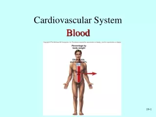
The cardiovascular system: blood. Chapter 11: Hematology. The functions of blood. Transportation : of dissolved gases, nutrients, hormones, and metabolic wastes. Regulation : of pH , blood clotting, body temperature, and renal control .

The cardiovascular system: blood
E N D
Presentation Transcript
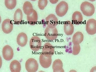
The cardiovascular system: blood Chapter 11: Hematology
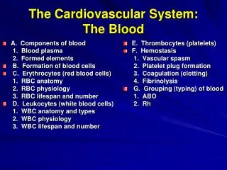
The functions of blood • Transportation: of dissolved gases, nutrients, hormones, and metabolic wastes. • Regulation: of • pH, • blood clotting, • body temperature, and • renal control. • Protection: against harmful toxins and pathogens
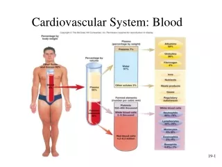
Anatomy of blood • Blood is a fluid connective tissue that contains: • Plasma (55%) • Dissolved proteins • Denser than water • Formed elements (45%) • Blood cells • Platelets (cell fragments)
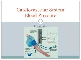
measuring blood • When we take blood, there are 3 characteristics we look at: • Temperature(100.4°F) • Viscosity (5x Water’s) • pH (7.35-7.45), Slightly Alkaline
plasma • Water takes up 92% of plasma volume • 8% Electrolytes, Proteins, Nutrients • Proteins: • Albumins (60%): of plasma proteins, produced in the liver, maintain osmotic balance (via water retention) • Globulins (35%): • Immunoglobulins: attack foreign proteins and pathogens; antibodies • Transport (escort) Proteins: carry compounds that are not water soluble (i.e. lipids) • Fibrinogens: function in blood clotting when converted to fibrin. *Liver synthesizes many of these proteins. Not this
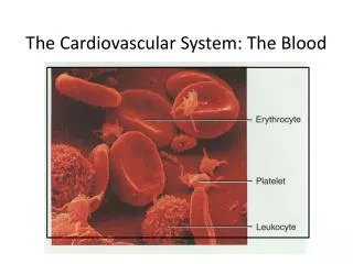
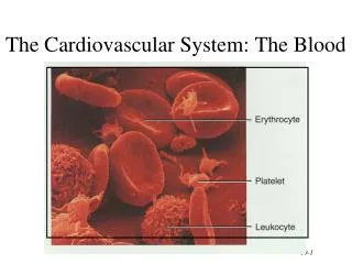
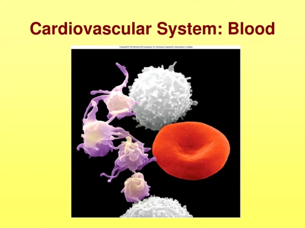
Formed elements • Produced via hemopoiesis (hematopoiesis) • Composition: (Hematocrit) • 99.9 % RedBloodCells (Erythrocytes) • <.1% WhiteBloodCells (Leukocytes) • ~ 1 to every 1000 RBCs • <.1% Platelets
Red blood cells • Shape: Biconcave discs • Mature RBCs lack nuclei and other organs (Erythropoiesis) • Life cycle: ~120 days; 1% replaced each day (3 million new cells each second!) • Hemoglobin: made of two globular proteins; contains heme pigment which holds an iron ion that binds with oxygen
Hemoglobin – iron in the blood heme a close up of Fe hemoglobin heme b
Clinical note: Sickle Cell ANemia • Cause: genetic mutation that affects the globular protein of hemoglobin • Sickled cells can easily clot and cause oxygen starvation of tissues
Antigens – can trigger an immune response • Antibody generator • Four Blood Types: • Type A: surface antigen A, plasma Anti-B Antibodies • Type B: surface antigen B, plasma Anti-A Antibodies • Type AB: surface antigen A & B, no anti-A or B antibodies • Type O: no surface antigens A or B, plasma Anti-A & Anti-B Antibodies • (+/-): Determined by presence or absence of Rh antigens on RBCs • Cross-reactions: mixing of blood types can cause agglutination (clumping) of RBCs and eventual hemolysis (RBC destruction) • Anti-Rh antibodies are not normally present in plasma, unless exposure sensitizes an individual.
Platelets – thrombocytes • Cell fragments that function in blood clotting • Formed from megakaryocytes • “Stick” together at injury sites to stop bleeding (hemostasis)
White blood cells – leukocytes • Larger than RBCs, have nuclei, lack hemoglobin • Function: defense against pathogens, toxins, wastes and abnormal/damaged cells • Two groups: granulocytes & agranulocytes • Amoeboid movement • Migration out of the blood stream • Some capable of phagocytosis
Granulocytes • Neutrophils • Eosinophils • Basophils • Agranulocytes • Monocytes • Lymphocytes
neutrophils • 70% of WBSs • First to arrive at an injury site • Active phagocytes (esp. bacteria) • Produced in bone marrow • 10-hour lifespan • Death recruits other WBCs and forms pus
eosinophils • 2-4% of WBSs • Two-lobed and dyed red by eosin • Attack anti-body labeled materials by exocytosis (secretion) of toxins • Produced in bone marrow
basophils • <1% of WBSs • Deep purple/blue with many granules • Migrate to injury sites and stimulates inflammation (via histamine) and prevents blood clotting (heparin) • Stimulated basophils release chemicals to attract other basophils and eosinophils • Produced in bone marrow
monocytes • 2-8% of WBSs • About twice as big as a RBC • Large free or fixed macrophages • Aggressive phagocytes • Active monocytes release chemicals to attract and stimulate other neutrophils, monocytes, phagocytes, and draw fibroblasts to the region. • Produced in bone marrow
lymphocytes • 20-30% of WBCs • Large nucleus with “halo” of cytoplasm • Integrates with lymphatic system • Do not rely on phagocytosis • Produced in bone marrow and lymphoid tissues
Abnormal amounts of leukocytes • Too few: leukopenia • Too many: leukocytosis • Extreme leukocytosis: leukemia • Cancer of blood-forming tissues.
hemostasis • Hemo “blood” + stasis “halt”: stops bleeding • Vascular Phase: constriction of damaged blood vessels (~30mins); blood vessel spasm • Platelet Phase: “sticky” platelets form a platelet plug • Coagulation Phase: conversion of fibrinogen to fibrin
Abnormal hemostasis • Excessivecoagulation • Thrombus: blood clot attached to wall • Embolus: moving blood clot • i.e. strokes, pulmonary embolism: immobile embolus within a blood vessel. • Inadequate coagulation • Hemophilia • Missing clotting factor • Symptoms including excessive bleeding (external and internal)