Cardiovascular System: Blood
270 likes | 291 Vues
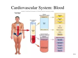
Cardiovascular System: Blood. Plasma. Liquid part of blood. Colloid : liquid containing suspended substances that don’t settle out of solution 91% water. Remainder proteins, ions, nutrients, waste products, gases, regulatory substances Proteins :

Cardiovascular System: Blood
E N D
Presentation Transcript
Plasma • Liquid part of blood. • Colloid: liquid containing suspended substances that don’t settle out of solution • 91% water. Remainder proteins, ions, nutrients, waste products, gases, regulatory substances • Proteins: • Albumins: viscosity, osmotic pressure, buffer, transports fatty acids, free bilirubin, thyroid hormones • Globulins: transports lipids, carbohydrates, hormones, ions, antibodies, and complement • Fibrinogen: blood clotting
Plasma, cont. • Ions: involved in osmosis, membrane potentials, and acid-base balance • Nutrients: glucose, amino acids, triacylglycerol, cholesterol, vitamins • Waste Products: • Urea, uric acid, creatinine, ammonia salts. Breakdown products of protein metabolism • Bilirubin. Breakdown product of RBCs • Lactic acid. End product of anaerobic respiration • Gases: oxygen, carbon dioxide, and inert nitrogen • Regulatory substances: hormones, enzymes
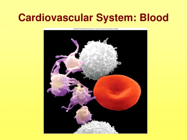
Formed Elements • Red blood cells (erythrocytes). Biconcave discs, anucleate, contain hemoglobin; transports oxygen and carbon dioxide. • White blood cells (leukocytes) • Granulocytes: cytoplasm contains large granules; have multi-lobed nuclei. Three distinctive types: neutrophils, eosinophils, basophils • Agranulocytes: cytoplasm contains small granules and nuclei that are not lobed. Two distinctive types: lymphocytes and monocytes • Platelets (thrombocytes). Cell fragment. Form platelet plugs, release chemicals necessary for blood clotting.
Red Blood Cells • Found in higher concentration in male than in female plasma • Components • 1/3 Hemoglobin • 2/3 Lipids, ATP, carbonic anhydrase
Hemoglobin Composition • Four globin molecules (polypeptide chains): Transport carbon dioxide (carbonic anhydrase involved), nitric oxide. NO brought from lungs to tissues, induces smooth muscles to relax, lowering BP. • Four heme molecules, each containing one iron atom: transport oxygen • Iron required for oxygen transport. Iron absorbed in upper small intestine; absorption increased by stomach acid and vitamin C. Iron lost in urine, feces, menstrual fluid.
Erythropoiesis • RBCs last 120 days in circulation (enucleated) • Production of red blood cells • Stem cells → proerythroblasts → early erythroblasts → intermediate erythroblasts → late erythroblasts → reticulocytes • Erythropoietin: hormone stimulates RBC production; produced by kidneys in response to low blood O2 levels.
Protect body against microorganisms and remove dead cells and debris Movements Ameboid: pseudopods Diapedesis: cells become thin, elongate and move either between or through endothelial cells of capillaries Chemotaxis: attraction to and movement toward foreign materials or damaged cells. Accumulation of dead white cells and bacteria is pus. White Blood Cells
Neutrophils: after leaving bone marrow, stay in circulation 10-12 hours then move into other tissues. Become motile, phagocytize bacteria, antigen-antibody complexes and other foreign matter. Secrete lysozyme. Last 1-2 days. Account for 60-70% of the WBC. • Eosinophils. Leave circulation and enter tissues during inflammatory response. Prevalent in allergic reactions. Destroy inflammatory chemicals like histamine. Release chemicals that help destroy tapeworms, flukes, pinworms, and hookworms. Account for 2-4% of the WBC.
Basophils: least common. Leave circulation and migrate through tissues, play a role in both inflammatory response and allergic reactions. Produce histamine and heparin. Account for less than 1% of the WBC. • Lymphocytes: produced in red bone marrow but then migrate to lymphatic tissues and proliferate. Responsible for antibody production. Studied extensively with the immune system. Account for 20-25% of the WBC. • Monocytes: remain in circulation for 3 days, leave circulation and become macrophages. Phagocytic cells. Can break down antigens and present them to lymphocytes for recognition. Account for 3-8% of the WBC.
Platelets • Cell fragments pinched off from megakaryocytes in red bone marrow • Important in preventing blood loss • Platelet plugs • Promoting formation and contraction of clots
Hemostasis • Arrest of bleeding • Events preventing excessive blood loss • Vascular spasm: Vasoconstriction of damaged blood vessels. Can occlude small vessels. Caused by thromboxanes from platelets and endothelin from damaged endothelial cells. • Platelet plug formation • Coagulation or blood clotting
Platelet Plug Formation • Platelet adhesion. Occurs when von Willebrand factor connects collagen and platelets. • Platelet release reaction. The release of ADP, thromboxanes, and other chemicals that activate other platelets. They in turn undergo the release reaction: cascade effect. • Platelet aggregation. Activated platelets express surface receptors that bind fibrinogen (protein found in plasma). Fibrinogen forms a bridge between platelets: platelet plug. • Expression of coagulation factor V and phospholipids. Important for coagulation
Coagulation • Stages • Activation of prothrombinase • Conversion of prothrombin to thrombin • Conversion of fibrinogen to fibrin • Coagulation factors. • Proteins found in plasma. • Circulate in inactive state until tissues are injured. • Damaged tissues and platelets produce chemicals that begin activation of the factors. • Pathways • Extrinsic • Intrinsic • Result: blood clot. A network of threadlike fibrin fibers, trapped blood cells, platelets and fluid
Blood Grouping • Transfusion: transfer of blood or blood components from one individual to another • Infusion: introduction of fluid other than blood • Determined by antigens (agglutinogens) on surface of RBCs • Antibodies (agglutinins) can bind to RBC antigens, resulting in agglutination (clumping) or hemolysis (rupture) of RBCs • Groups: ABO and Rh
Transfusion • Type A blood has anti-B antibodies; Type B blood has anti-A antibodies • Suggested that these antibodies are present because of exposure to A and B antigens on bacteria and food • Donor: gives blood. Recipient: receives blood • Type O as “universal donor”. Can actually cause transfusion reactions because of antibodies in O blood plasma
Rh Blood Group • First studied in rhesus monkeys • Types • Rh positive: Have these antigens present on surface of RBCs • Rh negative: Do not have these antigens present • Hemolytic disease of the newborn (HDN) • Rh positive fetus, Rh negative mother. • Late in pregnancy, Rh antigens of fetus cross placenta (through a tear in placenta or during delivery); mother creates antiRh antibodies (primary response) • Second Rh positive pregnancy might initiate secondary response and HDN (potentially fatal to fetus since antibodies to its RBCs would cross the placenta from the mother to the fetus, destroying fetal RBCs). • Injection of RhoGAM. Contains antibodies against Rh antigens. Antibodies attach to any fetal RBCs and they are destroyed.
Diagnostic Blood Tests • Type and Crossmatch: determination of ABO and Rh blood types. Red cells tested against antibodies • Complete Blood Count • Red Blood Count: number of RBCs/ microliter of blood • Hemoglobin Measurement: grams of hemoglobin/100 mL of blood. For a male, 14-18, female 12-16 g/100 mL • Hematocrit Measurement: percent of blood that is RBCs • White Blood Cell Count: 5,000-10,000 /microliter of blood
Differential White Blood Count: determines percentage of each of the five types of WBC • Neutrophils: 60-70% • Lymphocytes: 20-30% • Monocytes: 2-8% • Eosinophils: 1-4% • Basophils: 0.5-1% • Clotting • Platelet Count: 250,000- 400,000/microliter • Prothrombin TimeMeasurement: measures how long it takes for blood to start clotting. 9-12 seconds. To test, thromboplastin is added to whole plasma • Blood Chemistry: composition of materials dissolved or suspended in the plasma. Used to assess functioning of many body systems