Download
1 / 28
290 likes | 624 Vues
Cardiovascular System: Blood Vessels. Chapter 19. Blood Vessels. Closed system starting and ending at heart 3 major types Arteries carry blood away from the heart Systemic are O 2 rich Pulmonary are O 2 poor Capillaries have direct contact with tissues

E N D
Cardiovascular System:Blood Vessels Chapter 19
Blood Vessels • Closed system starting and ending at heart • 3 major types • Arteries carry blood away from the heart • Systemic are O2 rich • Pulmonary are O2 poor • Capillaries have direct contact with tissues • Veins carry blood toward the heart • Systemic are O2 poor • Pulmonary are O2 rich
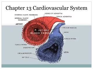
Blood Vessel Walls • 3 layers or tunics • Tunica intima • Simple squamous endothelia layer • Tunica media • Smooth muscle and elastin fibers • Sympathetic NS controls vasodilation and -constriction • Thicker in arteries to regulate pressure and flow • Tunica externa • Collagen fibers for support and reinforcement • Larger vessels with vasa vasorum • Central lumen contains the blood
Arteries • Elastic arteries are largest and nearest the heart • Large lumen why? • Tunica media w/ more elastin than smooth why? • Continuous, constant blood flow • Muscular arteries deliver blood to organs • Tunica media primarily smooth muscle why? • Arterioles lead to capillary beds • Tunica media scattered smooth muscle and little elastin • Vasodilation and –constriction alter capillary flow
Capillaries • Thin tunica intima w/basement membrane • Pericytes, similar to smooth muscle, to stabilize • Cells move single file • Accessible to most tissues for exchange • Types • Continuous abundant in skin and muscle • Endothelium uninterrupted • Tight junctions link cells • Fenestrated where rapid fluid exchange occurs • Endothelium with numerous pores • Pores thinly covered, but more permeable • Sinusoids in liver, bone marrow, and lymphoid tissue • Larger pores and lumens • Slower blood flow
Capillary Beds • Vascular shunts • Terminal arteriole • Metateriole • Thoroughfare channel • Postcapillaryvenule • True capillaries • Precapillaryspinchtersat junction with shunts • Rings of smooth muscle • Regulate flow based on chemical input and needs • Digestive tract: before and after a meal • Skeletal muscles: exercising and relaxing
Veins • Venules • Smallest are entirely endothelia and extremely porous • Larger with thin tunica media and tunica externa • Veins • 3 thinner tunicas • Media has little smooth muscle or elastin • Externa thickest • Larger lumens offer little blood flow resistance • Pressure lower • Hold ~65% of blood volume • Venous valves in limbs • Formed by tunica intima • Prevent blood backflow due to gravity
Vascular Anastomoses • Fusing of blood vessels in a given region • Provide multiple paths to/from organs/tissue • Near joints, also in heart and brain • Lacking in retina, kidneys, and spleen • Ensures uninterrupted blood flow • Metarteriole-thoroughfare channel is an example
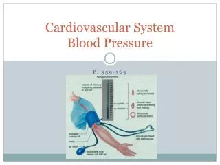
Physiology of Circulation Terminology • Blood flow (ml/min) • Amount of blood moving at a given time • Equivalent to cardiac output (CO) • Constant at rest; varies w/organ needs • Blood pressure (mm Hg) • Force exerted on a vascular wall • Systemic arteries used when measured • Keeps blood moving from high to low
Physiology of Circulation Terminology (cont.) • Peripheral resistance • Opposition to blood flow from vessel friction • Sources • Viscosity: thickness of blood • Changes in RBC numbers can increase/decrease, • Vessel length • Longer vessel = more resistance • More vessels = more total length • Vessel diameter • Smaller arterioles can constrict/enlarge • Flow is slowed along walls larger diameter = less wall contact • Turbulence, from additional wall resistance, increases
Basic Circulation Physiology • Blood flow = blood pressure/resistance SO • Increase pressure = increase flow BUT • Increase resistance = decrease flow • Resistance affects local flow more than pressure • Vasoconstriction/-dilation in an organ • Pressure basically unchanged overall
Systemic Blood Pressure • Highest at heart • Decreases w/increased distance • Steepest change in arterioles • Resistance highest • Flow maintained by pressure gradient
Arterial Pressure • Pressure near heart is pulsatile • Systolic pressure is max during ventricular contraction • Added blood supply stretches arteries • Diastolic pressure is minimum during ventricular relaxation • Recoil of arteries to maintain pressure • Difference creates pulse pressure, measured as our pulse • Increases w/ arteriosclerosis b/c elasticity decreases • Mean arterial pressure (MAP) is pressure moving blood to tissues • MAP = diastolic + 1/3 pulse pressure • Accounts for changes in arterial BP and longer diastole
Capillary Pressure • Significantly lower than arterial • Beneficial to capillary structure • Vessels are thin = fragile • Minimum needed to force filtrates out
Venous Pressure • Minimal changes in a cardiac cycle • Adaptations to compensate • Muscular pump: skeletal muscles • ‘Cankles’ and mats for standing • Respiratory pump: pressure created from inhalation • Valves • Smooth muscle in tunics
Monitoring Blood Pressure • Short-term • Cardiac output (CO) • Increase CO = increase BP • Decrease CO = decrease BP • See fig. 19.7 • Peripheral resistance (R) • Increase vasoconstriction = increase BP • Increase vasodilation = decrease BP • Long-term • Blood volume • Increase blood volume = increase BP • Decrease blood volume = decrease BP
Vasomotor Center • Monitors blood vessel diameter from medulla • SNS innervation (NE and ACh) • Minimizes moment specific changes to BP • Types of neural controls • Baroreceptors respond to vessel stretch • Carotid arteries and aortic arch • Inhibits vasomotor center vasodilation • Chemoreceptors respond to O2 and pH drop (CO2rise) • Close to baroreceptors • Stimulates vasomotor center vasoconstriction • Hypothalamus and cerebral cortex input to medulla
Hormonal Control of BP • Vasodilators • Atrial natriuretic peptide (ANP): increase Na+ and H2O excretion • Nitric oxide (NO): brief and localized • Inflammatory: histamines and prostocyclin • Alcohol: inhibits ADH • Vasoconstrictors • Adrenal medulla hormones: NE and Epi • Antidiuretic hormone (ADH): stimulates H2O conservation • Angiotensin II: renin from kidneys catalyzes production
Renal Regulation of BP • Monitors blood volume from kidneys • Increased BP stimulates H2O loss = decrease BP • Decreased BP stimulates H2O retention = increase BP • Excessive salt intake • Indirect mechanisms • Renin-angiotensin mechanism • Angiotensin II: vasoconstriction, aldosterone to resorb Na+/H2O, ADH release to resorb water • Direct mechanisms • Fluid speed increased to kidneys • Less absorption = more excretion • Figs 19.10 and 19.11
Measuring Circulation • Pulse • Measured at pressure points • Can be used to slow/halt distal blood flow • Blood pressure • Ausculatory method at brachial artery • First sound = systolic, no sounds = diastolic • Along with respiratory rate and body temp constitute vital signs
Blood Flow • Functions • Deliver O2 and nutrients; remove CO2 and wastes • Exchange gases • Absorb nutrients • Form urine • Rate dictated by needs (rest) • Brain (13%) • Heart (4%) • Kidneys (20%) • Abdominal organs (24%) • Skeletal muscles (20%) • All others (19%)
Blood Flow Velocity • Inversely related to cross-sectional area • More area (more vessels) = slower flow • Slowest in capillaries = more exchange time
Short-Tem Autoregulation • Adjustments of blood flow to tissue needs • Local, short-term intrinsic control • MAP and CO unchanged, but diameter of arterioles not • Metabolic controls (vasodilation) • Hb carries O2 and NO to tissues when O2 levels drop • K+, H+, adenosine, histamine too • Myogenic controls • Stretch receptors in smooth muscle • Less pressure signals vasodilation (how change flow?) • Fig 19.15
Long-Term Autoregulation • Angiogenesis increases blood vessel number and diameter • Short-term fails to meet needs • Occurs over weeks or months • Coronary occlusions or high-altitude living conditions
Capillary Diffusion • Via [gradient] • Nutrients and gases • Means vary based on molecule properties
Capillary Fluid Flow • Capillary hydrostatic pressure (HPc) force fluid out, more at arteriole than venule end • Capillary colloid osmotic pressure (OPc) draws fluid in, constant at both ends • Net filtration pressure (NFP) tells of net fluid loss or gain • NFP = (HPc – HPif) – (OPc – OPif) • More fluid enter tissue than return to blood, but lymphatic system returns
Circulatory Shock • Hypovolemic • From large loss of blood volume • HR up, vasoconstriction to increase venous return • Temporary fix; fluid replacement needed ASAP • Vascular • Extreme vasodilation drops resistance • Anaphylactic shock, septicemia, or ANS failure • Blood volume normal, but poor circulation • Cardiac • Heart fails
Blood Vessel Imbalances • Varicose veins: stretching veins due to leaky valves • Arteriosclerosis: hardening of arteries • Contributes to hypertension: BP > 140/90 mmHg • Most common is atherosclerosis: plaque build up of tunica intima • Hypotension: BP < 100/80 mm Hg • Less serious and often good • Can increase fainting/dizziness • Aneurysm: ballooning of a blood vessel • Phlebitis: inflammation of a vein