Cardiovascular System - Vascular System
Cardiovascular System - Vascular System. Blood & blood vessels. What are the components of blood?. ............... – Pale yellow, 90% Water, 8% Protein, 2% Salts ................................. – Iron rich Haemoglobin combines with oxygen & transported in blood


Cardiovascular System - Vascular System
E N D
Presentation Transcript
Blood & blood vessels What are the components of blood? ...............– Pale yellow, 90% Water, 8% Protein, 2% Salts ................................. – Iron rich Haemoglobin combines with oxygen & transported in blood ................................. – Fight infection and disease ..................... – Assist in clotting blood
Functions of blood What are the functions of blood?
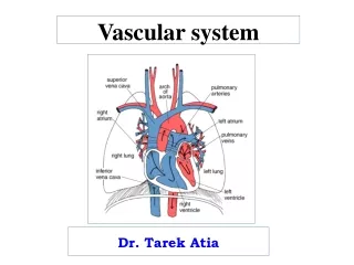
The Circulatory System This system is referred as a double circulatory system: • ................................... – blood between heart and lungs • ................................... – blood between heart and rest of the body
Blood Vessels • The vascular network through which blood flows to all parts of the body comprises of...................................... .............................................................................................................................................................................. & • Arteries are high pressure vessels which carry blood from the heart to the tissues. • The largest artery in the body is the aorta which is the main artery leaving the heart. • The aorta constantly subdivides and gets smaller. • The constant subdivision decreases the diameter of the vessel arteries, which now become arterioles.
Arteries Arteries are composed of three layers of tissue: • 1 an outer fibrous layer – • 2 a thick middle layer — • 3 a thin lining of cells to the inside — • The tunica media is comprised of smooth muscle and elastic tissue, which enables the arteries and arterioles to alter their diameter.
Arteries • Arteries tend to have more elastic tissue, while arterioles have greater amounts of smooth muscle; this allows the vessels to increase the diameter through vasodilation or decrease the diameter through vasoconstriction. • It is through vasoconstriction and vasodilation that the vessels can regulate blood pressure and ensure the tissues are receiving sufficient blood — particularly during exercise.
Arteries and arterioles have three basic functions: 1. .......................................................................... .......................................................................... 2. .......................................................................... .......................................................................... 3. .......................................................................... ..........................................................................
Veins and venules • Veins are .......................... vessels which return blood to the heart. The structure is similar to arteries, although they possess .............................. and elastic tissue. • Venules are the .............................. and transport blood away from the capillary bed into the veins. • Veins gradually increase in thickness the nearer to the heart they get, until they reach the largest vein in the body, the ............................, which enters the ........................... of the heart.
Veins and Venules • The thinner walls of the veins often distend and ........................................ in them. This is also allowed to happen as the veins contain .............................. which close intermittently to prevent .......................... of blood. • This explains why up to ................ of total blood volume is found in the venous system at any one time.
Capillaries • Capillaries are the functional units or the vascular system. • Composed of a ......................... of endothelial cells, they are just thin enough to allow ............................... to squeeze through their wall. • The capillary network is very well developed as they are so small; large quantities are able to cover the muscle, which ensures efficient .......................................
Capillaries • If the cross-sectional area of all the capillaries in a muscle cell were to be added together, the total area would be much greater than that of the aorta. • Distribution of blood through the capillary network is regulated by special structures known as pre-capillary sphincters, the structure of which will be dealt with later in this chapter.
Veins • Before looking at venous return it is important to look at the structure of veins. • Veins have thinner walls than arteries. • Veins also have valves.
Veins • The pressure of blood in the veins is too low to push blood back to the heart. • This problem is overcome in a number of ways. 1. 2. 3. 4. 5. 6. 7.
Skeletal Pump • The muscles surrounding the veins ...................................., pressing on veins and causing a pumping effect. • This muscle action is particularly important in maintaining .............. ......................................... • It is referred to as the ..............................
Adjacent Arteries • The surges of pressure in the adjacent arteries cause them to push against the veins, creating a regular pumping effect.
Pocket Valves • The blood in the veins can only move towards the heart; It cannot fall back to where it came from. • This is because at regular intervals there are ................................ ......................................situated in large veins.
Pocket Valves • These allow the free flow of blood towards the heart, but they close to prevent blood flow towards the heart, but they close to prevent ...................... ...................................
Inspiration • Increases thoracic volume, and so decreases thoracic pressure. • The vein in this region expand, causing blood to be ‘..............’ through them.
Gravity • ..........................the flow of venous blood from body parts above the heart. • However, ............................. the flow from parts below the heart.
Action of the Heart • The pumping action of the heart causes blood to flow in to replace the blood pumped out. • This creates a ................ ........................ in the veins close to the heart.
..................................... • There is an attraction between the molecules in any fluid moving in a particular direction, and this attraction helps maintain the constant flow. • This is called hydrostatic pressure. • This is important in blood, particularly as the fluid column moves back, ........................, to the heart.
Summary • The ........................ of blood in the veins is too ........ to push blood back to the heart. • This problem is overcome in a number of ways. • Hydrostatic Pressure • Gravity • Action of the Heart • Inspiration • Pocket Valves • Adjacent Arteries • Skeletal Pump
Blood Pressure • Blood pressure is the ................ exerted by the blood against the ............... of the blood vessels. • It is necessary to maintain ....................... through the circulatory system • It is determined by two main factors – • .............................. – the volume of blood flowing into the system from the left ventricle. • .............................. – the impedance offered by the blood vessels to the blood flow.
Blood Pressure • Blood pressure = ..................... X ...................... • Therefore, blood pressure increases when either cardiac output or resistance ......................... • Blood pressure in the .................. also increases and decrease in a pattern which corresponds to the cardiac cycle during .............................., when blood is pumped into the aorta and lowest during ............................
Blood Pressure Measurement • BP is usually measured as the brachial artery using a sphygmomanometer, and is recorded as mmHg of systolic pressure over diastolic pressure: • ..................... pressure is experienced when the heart pumps blood into the system • ..................... pressure is recorded when the heart is relaxing and filling with blood. • Typical Reading = 120mmHg 80mmHg
Blood Pressure and Exercise • BP Changes during exercise. • During Aerobic exercise, the systolic pressure ....................... as a result of increased cardiac output, while diastolic pressure .........................., or in well trained athletes may even drop as blood feeds into the working muscles. • During isometric or anaerobic exercise both systolic and diastolic pressure rise significantly due to increased resistance of the blood vessels.