Download
1 / 51
560 likes | 692 Vues
Explore the intricate structures of the heart wall and vascular system, from cardiac muscle to capillaries, with detailed explanations and key features.

E N D
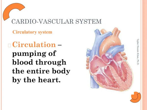
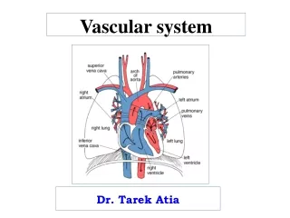
Vascular system Dr. Tarek Atia
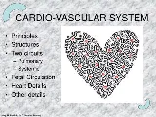
Wall of the heart • The wall of the heart contain: * A musculature of cardiac muscle * A fibrous skeleton * Impulse conducting system
Musculature of the heart • Epicardium: methothelial cells and its underlying CT, BV, and nerve fibers. • Myocardium: cardiac muscle fibers. • Endocardium: formed of endothelium and sub-endothelial CT.
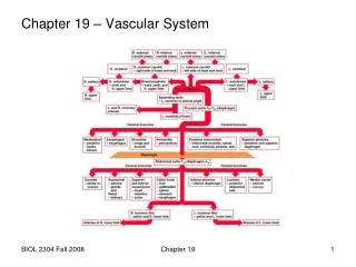
It Consists of 4 fibrous rings around the valve openings, and 2 fibrous trigones connecting the rings, and the membranous parts of the interventricular and interatrial septa. The fibers rings composed of dense irregular CT. Fibrous skeleton of the heart
Impulse conducting system • SAN • AVN • Bundle of Hiss • Rt bundle branch • Lt bundle branch • Purkinje fibers
1- Tunica Intima • Endothelium: single layer of Simple Squamous Epitheliumresting on Basal lamina • Subendothelial layer: loose CT, occasionally; contains some smooth muscle fibers. Sometimes; the subendothelial layer of arteries and arterioles contain a sheet of elastic fibers (internal elastic membrane).
2- Tunica Media • Circumferential layers of smooth muscle fibers, with collagen, elastic fibers and proteoglycanse in between. • Smooth muscle fibers secrete the fibers, and matrix. • Contraction of smooth muscles regulate and/or affect the blood pressure and blood flow. • Sometimes; a dense layer of fenestrated sheet of elastic fibers (external elastic membrane) separate tunica media from tunica adventitia.
3- Tunica Adventitia • The outer most CT layer composed of longitudinal bundles of collagen and elastic fibers; BV, lymphatics, and nerve supply.
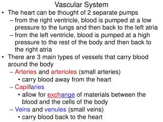
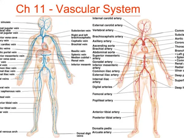
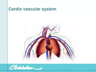
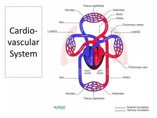
Types of BV • Large or elastic arteries • Medium sized or muscular arteries • Small arteries • Arterioles • Capillaries • Postcapillary venules • Small veins • Medium sized veins • Large veins • Atypical veins
Elastic or Large Arteries • Includes aorta, pulmonary arteries, and their main branched. They serve as conductor tubes. 1- Tunica intima • It is relatively thick. • Endothelium lining and its basal lamina. • Subendothelial layer: CTcontain collagen, elastic fibers, smooth muscle fibers; and macrophages. • Internal elastic membrane not well developed.
2- Tunica Media • It the most thick layer of the wall. • Formed of multiple layers of smooth muscle fibers separated by elastic lamellae. • Elastic lamellae are formed of fenestrated sheets of elastic fibers arranged in concentric layers.
3- Tunica adventitia • It is formed of Loose network of elastic and collage fibers. • It contain Fibroblasts, macrophages, blood vessels (vasa vasorum), and nerves (nervi vascularis).
Muscular, Medium Sized Arteries • Have more smooth muscle fibers and less elastic fibers in the tunica media
Tunica Intima: relatively thin, with well prominent Internal Elastic Membrane. • Tunica Media: Formed mainly of smooth muscle fibers, and few collagen and elastic fibers. • Tunica Adventitia: separated from tunica media by well prominent External Elastic Membrane. Formed of CT, collagen and elastic fibers, blood vessels, and nerves.
Capillaries • Have small diameter, less than the diameter of RBCs. • Have thin wall through it fluid containing gases, metabolites, and waste product can move. • Formed of single layer of endothelial cells, basal lamina, and sometimes Pericyes.
1- Continuous Capillaries • Found in the muscles, lung, and CNS. • Tight junctions appear to joint cells.
2- Fenestrated capillaries • Found in the endocrine glands, site of absorption as in intestine. • Contain fenestrations provide channels (with non-membranous diaphragm) across the wall.
3- Discontinuous capillaries • Sometimes called blood sinusoids. • Found in the liver, spleen and bone marrow. • They are large in diameter, and more irregular in shape. • Their structure vary between organs; but macrophages are associated within endothelial cells.
Veins • The tunics of veins are not well developed as those of arteries. • Veins have thinner wall than arteries. • The lumen of vein is large and collapsed. • Some veins contain valves, specially those of the lower limbs.
Medium sized veins • Tunica Intima: • Endothelial cells and basal lamina • Subendothelial CT, with some smooth muscle fibers. • A thin internal elastic membrane found in some veins. • Tunica media: • Thinner than tunica adventitia. • Many layers of smooth fibers, with collagen and elastic fibers in between. • Tunica Adventitia: • Thicker than tunica media • Contain collagen fibers, and a network of elastic fibers.
Medium sized veins Medium sized artery
Skin Dr. Tarek Atia
Introduction *Skinis the heaviest single organ of the body. * It is composed of : • Epidermis;Epithelium of ectodermal origin • Dermis; Layer of CT of mesodermal origin • Hypodermis; Or subcutaneous tissue; it is not a component of the skin.
Epidermis is composed of stratified squamous keratinized epithelium, containing keratinocytes and non-keratinocytes. • Dermis is dense irregular CT rich in Blood Vessels, collagen and elastic fibers.
Functions - It serves as a barrier against infection. - It is impermeable prevents pass of fluids from the external environment. - It is a hydrophobic layer prevents loss of body fluids. - It regulates body temperature by regulating the blood flow through the capillary beds and by sweating.
Types of skin Two types based on the comparative thickness of the epidermis into; thin and thick skin. • The epidermal layer of thin skin is about 75-150 µm, but it is about 400- 600µm in thick skin. • Thick skin is smooth, non-hairy, and is found in the palms and soles. • Thin skin is hairy, and is found elsewhere on the body.
Thick skin Thin skin
1- EPIDERMIS Epidermis contains two types of cells: A – Keratinocyts • They are keratin forming cells. • They are organized into layers and become mature in about 4 weeks. • Each layer represents a dynamic stage of cell division and cell maturation.
Stage of cell maturation • Cell renewal (Mitosis). • Cell differentiation (Keratinization) • Cell death • Exfoliation (Sloughing off the dead cells OFF)
B - Non-keratinocytes - Melanocytes: pigment cells - Langerhans cells: phagocytic cells - Merkel`s cells: sensory cells
Layers of Epidermis 1- Stratum Basale 2- Stratum spinosum 3- Stratum Granulosum 4- Stratum Lucidum 5- Stratum Corneum
1- Stratum Basale • Single layer of basophilic columnar or cuboidal cells resting on a basement membrane. • They contain stem cells (with high mitotic activity) responsible for renewal of epidermal cells. • All cells contain intermediate keratin filaments, the number of these filaments increases as cells progress upward.
2- Stratum Spinosum • It is composed of cuboidal or slightly flattened cells, with central nuclei. • The cytoplasm filled with bundles of keratin filaments (TONOFILAMENTS) • Cells of this layer are firmly bound together by the filaments filled the cytoplasmic spines and desmosomes that punctate the cell surface, giving them spine-like appearance.
3- Stratum Granulosum • It is composed of 3-5 layers of flattened polygonal cells, whose cytoplasm is filled with non-membranous basophilic granules (KeratohyalinGranules) and membranous granules (Lamellar granules). • The lamellar granules contain Lamellar Disks of Lipid Bilayers. • These granules fuse with the cell membrane and release their lipid content into the intercellular spaces forming a Sheet of Lipid Deposit, which acts as a barrier sealing the skin.
4- Stratum Lucidum - More common in thick skin, as a translucent thin layer of flattened eosinophilic non-nucleated cells. - The cytoplasm consists of densely packed Keratin Filamints embedded in electron dense matrix, but all cell organoids are disappeared.
5- Stratum Corneum • It composed of 15-20 layers of flattened non-nucleated keratinized cells (horny cells) , their cytoplasm is filled with Keratin
1- Melanocytes • Melanocytes have long cytoplasmic processes that branch into the epidermis, running between cells of stratum basale and spinosum. • Melanocytes present normally beneath or between cells of stratum basale. • There are two types of melanin: • Eumelanin: dark brown pigment produced by melanocytes. • Pheomelanin: reddish pigment present in red hair.
Mechanism of melanin formation • Melanin synthesis starts in rER; then it is concentrated, accumulated and stored in Golgy bodies. • The pigments are found in secretory vesicles called Melanosomes. • Then the pigment granules leave melanocytes and phagocytosed by keratinocytes.
2- Langerhans Cells • They are star shaped cells, with indented nuclei. • They are phagocytic skin cells present within the cells of Stratum Spinosum; and represent 2- 8% of the cells. • The cytoplasm contain special granules called BIRBECKS GRANULES.
3- Merkel’s Cells - Sensory skin cells. - Located mainly in thick skin. - Act as mechanoreceptors.
Dermis • It is a CT layer rich in BV, lymphatics, and CT cells. • It is formed of two layers:- • Papillary layer • Reticular layer