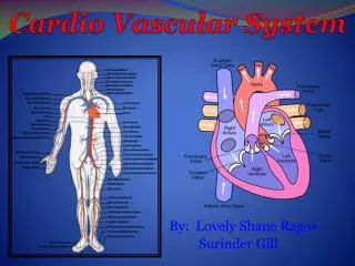
Vascular System
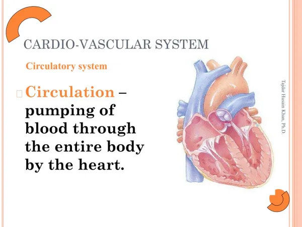
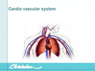
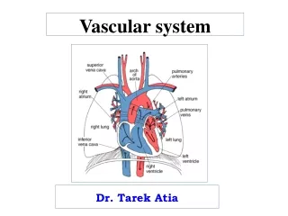
Vascular System. The heart can be thought of 2 separate pumps from the right ventricle, blood is pumped at a low pressure to the lungs and then back to the left atria from the left ventricle, blood is pumped at a high pressure to the rest of the body and then back to the right atria

Vascular System
E N D
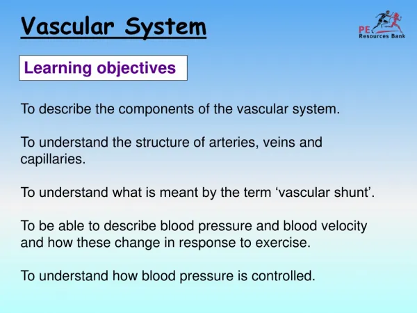
Presentation Transcript
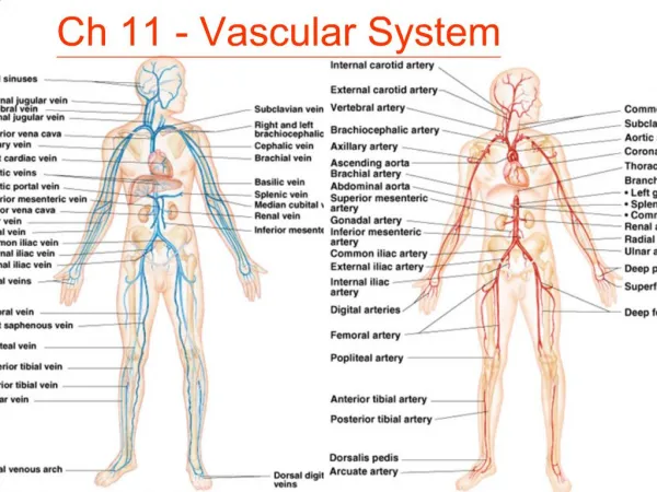
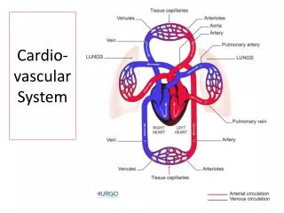
Vascular System The heart can be thought of 2 separate pumps from the right ventricle, blood is pumped at a low pressure to the lungs and then back to the left atria from the left ventricle, blood is pumped at a high pressure to the rest of the body and then back to the right atria There are 3 main types of vessels that carry blood around the body Arteries and arterioles (small arteries) carry blood away from the heart Capillaries allow for exchange of materials between the blood and the cells of the body Veins and venules (small veins) carry blood back to the heart
Vascular Pathways Arteries and arterioles are characterized by a divergent pattern of blood flow blood leaves each ventricle via a single artery but split into numerous and smaller diameter vessels Arterioles branch into capillaries capillaries are the most numerous blood vessel with the smallest diameter Venules and veins are characterized by a convergent pattern of blood flow blood flows out of many capillaries into a single venule with a larger diameter from the venules, blood flows into veins that are larger in diameter which merge into a single vessel to deliver blood to the atria ~60% of the blood volume at rest is in the veins
Vascular Walls All blood vessels are lined with a thin layer of endothelium, a type of epithelium which is supported by a basement membrane called the tunica intima (or tunica interna) only layer of capillary walls The walls of most arteries and veins have layers of smooth muscle and/or elastic connective tissue called the tunica media and fibrous connective tissue called the tunica externa, surrounding the endothelium the thickness of the tunica media and externa vary in different vessels depending on their function or the amount of internal (blood) pressure that they encounter
Smooth Muscle Most blood vessels contain vascular smooth muscle arranged in circular layers which is partially contracted at all times creating a condition known as muscle tone Additional contraction of the smooth muscle results in vasoconstriction which narrows the diameter of the vessel lumen Relaxation of the smooth muscle results in vasodilation which widens the diameter of the vessel lumen Neurotransmitters, hormones and paracrine signals influence vascular smooth muscle tone which in turn will affect blood pressure and blood flow throughout the cardiovascular system
Blood Flow Through Vascular System Totalblood flow through any level of the circulation is equal to the cardiac output if cardiac output is 5 L/min, the blood flow through all systemic capillaries is also 5 L/min blood flow through the pulmonary side is equal to blood flow through the systemic circulation prevents blood from accumulating in either the systemic or pulmonary loop
Distribution of Blood Flow The distribution of systemic blood varies according to the metabolic needs of individual organs and is governed by homeostatic reflexes skeletal muscles at rest receive 21% of cardiac output, but during exercise when they use more O2 and nutrients and produce more CO2 and wastes receive as much as 85% of cardiac output accomplished through the vasoconstriction and vasodilation of arterioles supplying blood to various regions, organs or tissues of the body The ability to selectively alter blood flow to organs is an important aspect of cardiovascular regulation
What Determines Blood Flow? Blood flow (F) through the vascular system is directly proportional to the pressure gradient (ΔP) between to points within the system: F ΔP if the pressure gradient increases, flow increases if the pressure gradient decreases, flow decreases blood pressure is the amount of force blood exerts outwardly on the wall of a vessel The tendency of the vascular system to oppose blood flow is called its resistance (R) and is inversely proportional to flow: F 1/R if the resistance increases, flow decreases if the resistance decreases, flow increases Combining the equations above results in: F ΔP/R
Blood Pressure Aortic pressure reaches an average high of 120 mmHg during ventricular systole (systolic pressure) and falls steadily to a low of 80 mmHg during ventricular diastole (diastolic pressure) systolic pressure > 120 is called hypertension systolic pressure < 100 is called hypotension The highly elastic walls of the arteries allows them to capture and store the energy of ventricular ejection note that the pressure in the aorta drops only to 80 mmHg (not to 0mmHg as observed in the ventricle) which keeps blood constantly moving (never stops) energy stored by the arteries can be felt as a pulse Blood pressure decreases as it flows downstream A similar blood pressure profile (albeit lower) is observed on the pulmonary side of circulation
What Determines Arterial BP? Arterial blood pressure is directly proportional to the amount of blood found in an artery more blood in an artery = higher pressure less blood in an artery = lower pressure Since arterial pressure is pulsatile, the mean arterial pressure (MAP) is used to represent the driving pressure of blood through the vascular system MAP = diastolic + 1/3 (systolic – diastolic) MAP = 80 + 1/3 (120 – 80) = 93 mmHg in the aorta
Mean arterial pressure is a balance between blood flow into the arteries and blood flow out of the arteries • if flow in exceeds flow out, pressure increases • if flow out exceeds flow in, pressure decreases • Blood flow in is equal to the cardiac output • Blood flow out is influenced primarily by the vascular resistance offered by the arterioles determined mainly by their diameter • MAP CO X Resistancearterioles
Regulation of Mean Arterial Blood Pressure The central nervous system coordinates the reflex control of blood pressure The main integrating center is a cluster of neurons in the medulla oblongata called the cardiovascular control center Sensory input to the integrating center comes from a variety of peripheral sensory receptors stretch sensitive mechanoreceptors known as baroreceptors in the walls of the aorta and carotid arteries travel to the cardiovascular center via sensory neurons Responses by the cardiovascular center is carried via both sympathetic and parasympathetic neurons and include changes in cardiac output and peripheral resistance which occur within 2 heartbeats of the stimulus
Baroreceptor Reflex The baroreceptors are tonically active stretch receptors that fire action potentials continuously at normal blood pressures When blood pressure increases in the arteries stretches the baroreceptor cell membrane, the firing rate of the receptor increases in response, the cardiovascular center increases parasympathetic activity and decrease sympathetic activity to slow down the heart decreased sympathetic outflow to arterioles causes dilation allowing more blood to flow out of the arteries When blood pressure decreases in the arteries, the cardiovascular center increases sympathetic activity and decreases parasympathetic activity creating opposite responses in the effectors to increase blood pressure
What Else Determines Mean Arterial BP? Although the volume of blood is usually relatively constant, changes in blood volume can affect mean arterial blood pressure if blood volume increases, blood pressure increases fluid intake if blood volume decreases, blood pressure decreases fluid loss Relative distribution of blood between the venous and arterial sides of circulation is an important factor in regulating arterial blood pressure when arterial blood pressure falls, vasoconstriction of the veins redistributes blood to the arterial side
Systemic Venous Blood Pressure As blood moves through the vessels, pressure is lost due to friction between the blood and the vessel walls The low pressure blood in veins inferior to the heart (arms, abdominopelvic cavity and legs) must flow against gravity to return to the heart To assist venous flow, these veins have internal one way valves to ensure that blood passing the valve cannot flow backward The movement of blood through veins is also assisted by the contraction ofskeletal muscle Veins located between skeletal muscles are squeezed during contraction This increases the venous pressure enough to move the blood through the valves, back towards the heart
What Determines Resistance in the Vessels? For fluid flowing through a tube, resistance is influenced by 3 parameters: the radius (r) of the tube (half of the diameter) the length (L) of the tube the viscosity (η) or thickness of the fluid Poiseuille’s Law relates these factors to resistance: R Lη/r4 if the tube length increases, resistance increases if the viscosity increases, resistance increases if the tube’s radius increases, resistance decreases Since blood viscosity remains relatively constant and blood vessel lengths can’t change, vessel diameter is the major determinant of resistance
Arteriolar constriction reduces blood flow through that arteriole and redirects the flow through all arterioles with a lower resistance • total blood flow through all the arterioles of the body always equals cardiac output
Local and Systemic Control of Arteriolar Diameter Local control is accomplished by paracrines secreted by the vascular endothelium or by tissues to which the arterioles are supplying blood low O2 and high CO2 dilate arterioles which increase blood flow into the tissue bringing additional O2 while removing excess CO2 can be caused by an increase in metabolic activity (active hyperemia) or by a period of low perfusion (reactive hyperemia) Systemic control occurs by sympathetic innervation tonic release of norepinephrine which binds to α-adrenergic receptors on vascular smooth muscle helps maintain tone of arterioles if sympathetic release of norepinephrine decreases, the arterioles dilate, if the release of norepinephrine increases, arterioles constrict
Capillary Wall Promotes Exchange Most cells are located within 0.1 mm of the nearest capillary over which diffusion occurs rapidly The most common type are continuous capillaries endothelial cells are joined by leaky junctions Less common type are fenestrated capillaries endothelial cells have large pores (fenestrations) that allow high volumes of fluid to pass quickly between the plasma and interstitial fluid Exchange occurs either by: movement of substances through the gaps between adjacent endothelial cells (paracellular movement) movement of substances through/across the cell membrane of endothelial cells (transcellular movement)
Capillary Exchange Paracellular exchange occurs through endothelial cell junctions or fenestrations solutes can move by diffusion solutes can move by bulk flow which refers to the mass movement of a solvent as a net result of hydrostatic and or osmotic pressure gradients across the capillary wall if the direction of bulk flow is out of the capillary the fluid movement is called filtration if the direction of bulk flow is into the capillary the fluid movement is called absorption Transcellular exchange occurs through the cell membrane of endothelial cells nonpolar gasses and solutes can move by diffusion large polar solutes can move by vesicular transport
Capillary Exchange by Bulk Flow 2 forces regulate bulk flow in capillaries hydrostatic pressure (Pcap) lateral pressure component of blood flow that pushes plasma out through the capillary pores decreases along the length of the capillary as energy is lost to friction osmotic pressure (pcap) pressure exerted by solutes within the plasma the main solute difference between plasma and interstitial fluid is due to proteins (present in plasma, but mostly absent in interstitial fluid) the osmotic pressure created by plasma proteins is called colloid osmotic pressure favors water movement by osmosis from interstitial fluid into plasma is constant along the length of the capillary
Net Pressure = Pcap – pcap Net Pressurearterial end = 32mmHg – 25mmHg = 7mmHg favors filtration Net Pressurevenous end = 15mmHg – 25mmHg = -10mmHg favors absorption
In most capillaries there is more filtration than absorption 90% the volume of fluid filtered out at the arterial end is absorbed back into the capillary at the venous end the other 10% enters lymphatic vessels where it is returned back into circulation as the lymph vessels empty lymph fluid into blood at the right atrium