Understanding Vascular System: Functions & Structures
Learn about arteries, capillaries, veins layers, and functions within the vascular system. Understand the different types of capillaries and their roles in circulation pathways.

Understanding Vascular System: Functions & Structures
E N D
Presentation Transcript
Chapter 19 – Vascular System Chapter 19
A. categories and general functions: • 1. arteries - carry blood • away from heart • 2. capillaries - allow • exchange of materials • between blood and • tissue fluid • 3. veins - return blood • to heart Chapter 19
B. wall structure • most vessel walls have 3 layers • lumen = space inside vessel • 1. tunica intima / tunica interna • endothelium – • simple squamous e. • subendothelial layer – • loose c.t. (collagen) Chapter 19
2. tunica media • a. smooth muscle - cells circularly arranged • controlled by ANS and chemical factors • constriction decreases blood flow and increases systemic blood pressure • dilation increases blood flow and decreases systemic blood pressure • b. elastic c.t. Chapter 19
3. tunica adventitia / tunica externa • c.t. attaches vessel to surrounding structures • vasa vasorum nourish outer part of vessel wall Chapter 19
C. arteries • 1. elastic (conducting) - large arteries near heart (aorta and major branches) • * conduct blood to muscular arteries • * low resistance • * tunica media = circular elastin sheets with few smooth m. cells • * recoil maintains blood pressure during diastole Chapter 19
2. muscular - middle-sized arteries, distal to elastic arteries • * distal to elastic arteries • * tunica media very thick; much smooth m. and some elastin • * regulate blood flow to organs • * have an internal and an external elastic lamina Chapter 19
3. arterioles - smallest arteries • * tunica media contains smooth m. only • * diameter controlled by ANS and chemical messengers • * diameter determines blood flow and blood pressure Chapter 19
D. capillaries • * wall consists of endothelium and basal lamina (no tunica media or externa) • * 8 to 10 mm in diameter • * join and branch to form capillary beds • * cells are joined at spots around perimeter by tight junctions and desmosomes • * intercellular clefts are spaces between cells Chapter 19
1. types • a. fenestrated capillaries (high permeability) • * have fenestra (openings) in endothelial cells • * some fenestra are covered by a membrane, others are not • * also have intercellular clefts • * found in small intestine, synovial joints, kidney Chapter 19
b. continuous capillaries • * intercellular clefts but no fenestra • * most common type • c. sinusoids • * wide, leaky capillaries, usually fenestrated • * fewer cell junctions • * allow passage of large particles • * found in bone marrow, spleen, liver Chapter 19
d. low-permeability capillaries • * complete tight junctions • * no fenestra, no clefts • * restricted transport vesicles • * can transport specific molecules in or out • * found in brain (blood-brain barrier) Chapter 19
2. capillary beds • precapillary sphincter - smooth m. cell wrapped around origin of capillary • * controls blood flow through capillaries • * sphincter controlled by autoregulation • * when sphincter is closed, blood is diverted to thoroughfare channel Chapter 19
E. veins • have thinner walls than arteries for the same diameter (larger lumen) • act as capacitance vessels - store extra blood (65%) • low pressure • tunica adventitia thicker than tunica media • venous valves prevent backflow Chapter 19
F. vascular anastomosis • occurs when vessels join midstream • anastomoses provide alternate pathways (collateral channels) Chapter 19
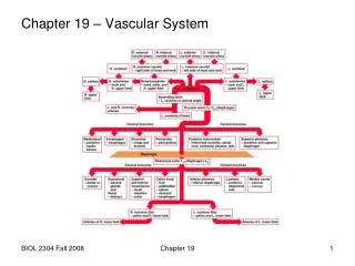
G. circulatory routes • 1. pulmonary circuit: right ventricle to lungs to left atrium • * pressure supplied by right ventricle • * low pressure system • * takes low oxygen blood (75% saturated) to lungs and brings high oxygen (98% • saturated) blood • back to heart Chapter 19
right ventricle • pulmonary semilunar valve • pulmonary trunk • left and right pulmonary arteries • lobar arteries (3 R, 2 L) take blood to lung lobes • pulmonary capillaries • pulmonary veins (superior and inferior, L and R) • left atrium Chapter 19
2. systemic circuit: left ventricle to body to right atrium • * pressure supplied by left ventricle • * high pressure system • * takes O2 to tissues and removes CO2 • * distributes nutrients from digestive tract to body • * collects wastes and takes them to kidney for excretion Chapter 19
a. circulatory pathways of the brain (cerebral arterial circle, circle of Willis) Chapter 19
anterior communicating a. • anterior cerebral a. • internal carotid a. • posterior communicating a. • posterior cerebral a. • basilar a. • vertebral a. Chapter 19
cross section of neck: Chapter 19
b. hepatic portal circulation • portal system = two capillary beds in series, joined by veins or arteries Chapter 19
capillaries of small intestine, part of large intestine and stomach • superior mesenteric vein • capillaries of spleen, stomach and pancreas • splenic vein • capillaries of distal large intestine and rectum • inferior mesenteric vein Chapter 19
hepatic portal vein • liver • liver sinusoids • hepatic veins • inferior vena cava Chapter 19
c. fetal circulation • placenta = organ formed from extra-embryonic membranes and endometrium to exchange materials between fetal and maternal blood • the umbilical veins take high-oxygen and high-nutrient blood from the placenta to the fetus • the umbilical arteries take low-oxygen, high-waste blood back to the placenta Chapter 19
umbilical vein from placenta • fetal liver • hepatic portal vein • ductus venosus • inferior vena cava • right atrium foramen ovale left atrium • right ventricle left ventricle • pulmonary trunk ductus arteriosus aorta • lungs • internal iliac a. • umbilical a. to placenta Chapter 19
adaptations: • 1) to bypass the fetal liver • ductus venosus - allows some umbilical vein blood to go through the fetal liver but diverts most of it directly into the inferior vena cava • 2) to bypass the non-functional fetal lungs • foramen ovale – opening in interatrial septum that allows blood to go from the right atrium directly into the left atrium • ductus arteriosus – vessel that connects pulmonary trunk and aorta Chapter 19