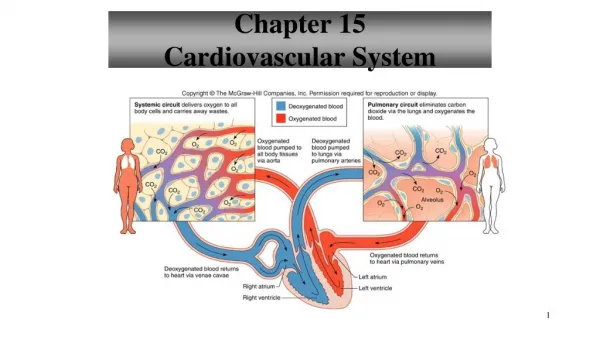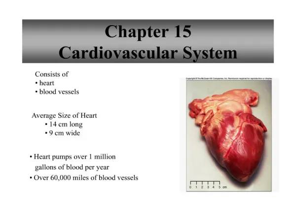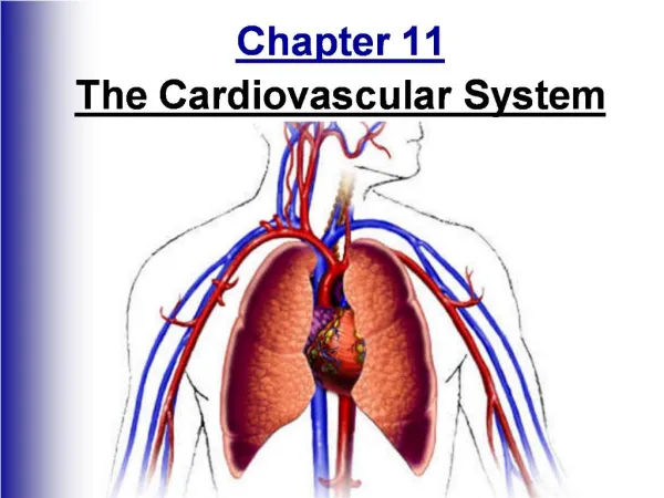
Chapter 13 Cardiovascular System
Chapter 13 Cardiovascular System. p. 357-363 Blood Vessels. Blood Vessels. Forms closed circuit that carries blood from the heart to cells and back Includes: Arteries Arterioles Capillaries Venules Veins. Arteries and Arterioles. Arteries

Chapter 13 Cardiovascular System
E N D
Presentation Transcript
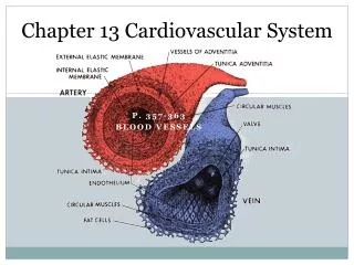
Chapter 13 Cardiovascular System p. 357-363 Blood Vessels
Blood Vessels • Forms closed circuit that carries blood from the heart to cells and back • Includes: • Arteries • Arterioles • Capillaries • Venules • Veins
Arteries and Arterioles • Arteries • Are strong, elastic vessels adapted to carry blood away from heart at high pressures • Arterioles • Fine branches of arteries
Arterial Wall • 3 layers • Endothelium (Tunica interna) • Helps prevent blood clotting • Helps regulate local blood flow by secreting substances that dilate or constrict blood vessels • Inner layer • Middle Layer (Tunica Media) • Makes up bulk of wall • Smooth muscle • Outer Layer (Tunica externa) • Attaches artery to surrounding tissues • Arterioles • Same three layers except much thinner and lead to capillaries
Constriction and Dilation • Vasoconstriction • Artery walls reduce diameter by muscle contraction • Vasodilation • Relaxing of muscles causes increase in diameter
Atherosclerosis • Nearly half of all deaths in the U.S. are due to this arterial disease • Occurs when soft masses of fatty materials, particularly cholesterol, accumulate on the inner surface of arterial walls • Known as plaque • Can cause blood clot formation which leads to a thrombus (blood clot) or embolus (lodging of clot somewhere) or even blood deficiency (ischemia) or tissue death (necrosis)
Atherosclerosis Cont. • Risk factors • Fatty diet, elevated blood pressure, smoking, obesity, and lack of physical exercise • Emotional and genetic factors may also increase susceptibility • Three common treatments • Percutaneoustransluminal angioplasty (balloon) • Laser angioplasty • Bypass graft surgery (triple and quad bypass)
Capillaries • Smallest diameter of vessels that exchange substances between blood and tissues • Slits • Openings in walls of capillaries • Size depends on type of tissue they are in • Capillary density • Depends on tissues rate of metabolism • Tissues with large quantities of oxygen and nutrients are packed with capillaries • Slow metabolic rates lack capillaries (cornea, epidermis)
Exchanges in Capillaries • Pre-capillary sphincters open and close capillaries • Also function to route blood flow • When exercising more blood is needed by skeletal muscles and less in digestive tract or other areas • Gases, nutrients, and metabolic by-products are exchanged between blood in capillaries and tissue fluids
Diffusion, Osmosis and Filtration • Diffusion • High concentrations of oxygen and nutrients entering, substances diffuse through capillary walls to tissues where blood meets CO2 and waste • Filtration • Forces molecules through membrane with hydrostatic pressure. When ventricle walls contract it provides force for filtration. • Osmosis • Plasma proteins that remain in capillaries makes osmotic pressure of blood greater than tissue fluid
D, O, F continued Blood pressure decreases with distance from heart because friction slows flow Normally more fluid leaves capillaries than what enters Unusual events may increase blood flow to capillaries and excess fluid enters spaces between tissue cells.
Veins and Venules • Venules • Microscopic vessels that continue from the capillaries to veins • Veins • Carry blood back to atria, roughly parallel to arteries • Walls are similar to arteries but much weaker • Function as blood reservoirs • If hemorrhage cause a drop of blood pressure, veins receive impulse to send more blood to heart to help maintain pressure. • Flapvalves • In upper and lower limbs • Help prevent blood from backing up by closing • Valves are open if blood is flowing toward heart but close if going in other direction • Table 13.2 summarizes vessels
Important to Remember • Arteries carry oxygenated blood away from heart • Veins carry deoxygenated blood towards heart • Exception • Pulmonary artery and pulmonary vein are opposite of above statement
Review Describe the wall of an artery. What is the function of smooth muscle in arterial wall? How is the structure of an arteriole different from that of an artery? Describe the capillary wall. What is the function of a capillary? What structures control blood flow into capillaries? What forces cause the exchange of substances between blood and tissue fluid? Why is the fluid movement out of a capillary greater at the capillary’s arteriolar end than at its venule end? How does the structure of a vein differ from that of an artery? How does venous circulation help to maintain blood pressure when hemorrhaging cause blood loss?