Download
1 / 83
910 likes | 1.73k Vues
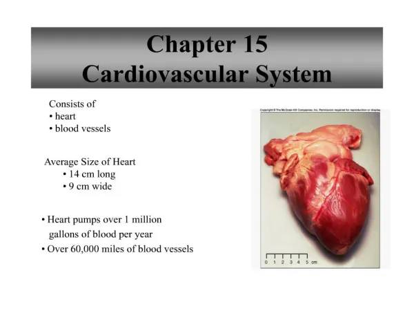
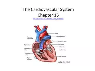
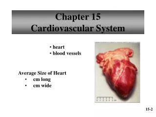
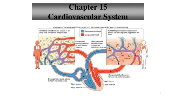
Chapter 15 Cardiovascular System. Structure of the Heart Hollow, cone shaped, muscular pump Size and Location of the Heart Average size about 14cm long and 9cm wide Bordered laterally by the lungs, posteriorly by the spinal cord, and anteriorly by the sternun

E N D
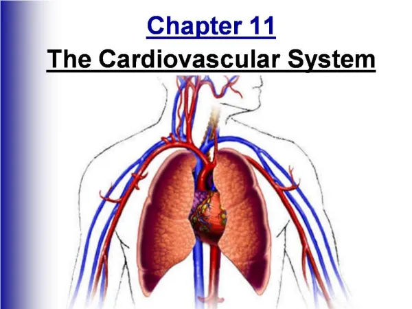
Structure of the Heart • Hollow, cone shaped, muscular pump Size and Location of the Heart • Average size about 14cm long and 9cm wide • Bordered laterally by the lungs, posteriorly by the spinal cord, and anteriorly by the sternun • Distal end extends downward and to the left (apex)
Size of Heart Average Size of Heart • 14 cm long • 9 cm wide
Coverings of the Heart • Pericardium – outer fibrous bag that encloses heart and the proximal ends of large blood vessels attached to heart - surrounds a more delicate double layered sac - inner layer (epicardium) covers the heart
Wall of the Heart • Epicardium – protective outer covering that secretes • Serous fluid • Myocardium - middle layer made mostly of muscle that • pumps blood out of the heart chambers • Endocardium – inner layer that lines all heart chambers • and covers the valves
Heart Chambers and Valves • 4 hollow chambers (2 left and 2 right) • Upper chambers (atria) are thin walled and receive blood returning to the heart • Lower chambers (ventricles) are more muscular and force blood out of the heart into arteries • Valves of the Heart -tricuspid, pulmonary, bicuspid (mitral), aortic – Table 15.2 Page 561
Mitral valve prolapse (Page 558) – mitral valve stretches and bulges into left atrium during contraction causing blood to sometimes regurgitate into left atrium • Chordae tendinae are tough fibrous strings attached to papillary muscle that prevent tricuspid and bicuspid valves from swinging back into atrium
Heart Valves Tricuspid Valve Pulmonary and Aortic Valve
Skeleton of Heart • fibrous rings to which the heart valves are attached
Blood Supply to the Heart • First two branches of the aorta (coronary arteries) supply blood to the tissues of the heart – Fig 15.13 Page 565 • Branches of the coronary veins drain blood from heart tissues – they join the coronary sinus and empty into the right atrium
Heart Actions • Cardiac Cycle = atria contract while ventricles relax, ventricles contract while atria relax, atria and ventricles relax for a moment
Heart Actions Atrial Diastole/Ventricular Systole Atrial Systole/Ventricular Diastole
Cardiac Cycle • A-V valves = atrioventricular valves (tricuspid and bicuspid) • During cycle pressure within chambers rises and falls • Pressure increases as chambers fill • Blood flows into atria when relaxed, A-V valves open when pressure is greater than in ventricles
Atria contract forcing remaining blood into ventricles (30%) • Atrial relaxation follows (diastole) • As ventricles contract A-V valves close (systole) • Pulmonary and aortic valves open and blood is ejected into arteries (aorta or pulmonary) • When ventricles relax A-V valves open and ventricles begin to fill with blood again
Heart Sounds • Through a stethescope it sounds like lubb – dupp • Blood flow through chambers and opening and closing of valves causes vibrations • Lubb sound occurs during ventricular contraction (systole) when A-V (bicuspid & tricuspid) valves are closing • Dupp sound occurs during ventricular relaxtion (diastole) when pulmonary and aortic valves are closing
Heart sounds can indicate condition of heart valves condition Ex: valve cusps may not close completely causing a murmur – many are harmless, most serious problems can be repaired or the valve replaced • Can hear heart sounds associated with each valve – fig 15.18 page 569
Cardiac Muscle Fibers • A functional syncytium is a mass a of merging cells that act a unit • Heart has 2 units • atrial syncytium – walls of atria • ventricular syncytium- wall of ventricles • units are connected by fibers of the cardiac conduction system in a small area of right atrial floor
Cardiac Conduction System S-A node • small mass of cardiac muscle tissue beneath the epicardium that initiate and distribute impulses throughout the myocardium • can reach threshold on its own without stimulation from nerve fibers • generates the heart’s rhythmic activity (70-80 times / min) • often called the pacemaker • right and left atria contract almost simultaneously
A-V node • Impulses from S-A node pass slowly to A-V node • Impulses then travel rapidly along A-V bundle and Purkinje fibers stimulating ventricles to contract • See fig 15.19 page 570 – locations • See fig 15.20 page 570 – pathway
Electrocardiogram (ECG) • Recording of electrical changes that occur in the myocardium during a cardiac cycle • Used to assess heart’s ability to conduct impulses • Electrodes are placed on the skin to record • Normal ECG fig 15.22 page 572 • P wave – deplolarization (contract) of atria • QRS complex – depolarization of ventricles • T wave – repolarization (relax) of ventricles
Clinical Application Arrhythmias Ventricular fibrillation • rapid, uncoordinated depolarization of ventricles Tachycardia • rapid heartbeat Atrial flutter • rapid rate of atrial depolarization
Regulation of Cardiac Cycle • Physical exercise and elevated body temperature increase heart action • Vagus nerves (parasympathetic fibers) secrete Ach • which decrease S-A and A-V activity – heart rate decreases • K ions affect the electrical potential of cell membranes • Ca ions needed for muscle contraction • Clinical application 15.2 page 574 – arrhythmias
Regulation of Cardiac Cycle Autonomic nerve impulses alter the activities of the S-A and A-V nodes
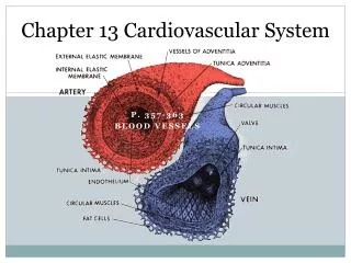
Blood Vessels • Table 15.3 Page 581- characteristics of blood vessels • Fig 15.25 - walls of an artery and vein • 15.26 Page 576 – walls of arterioles • fig 15.29 page 578 walls of capillaries
Arteriole • smallest arterioles only have a few smooth muscle fibers • capillaries lack muscle fibers
Metarteriole connects arteriole directly to venule
Capillaries • smallest diameter blood vessels • extensions of inner lining of arterioles • walls are endothelium only • semipermeable • sinusoids – leaky capillaries
Regulation of Capillary Blood Flow Precapillary sphincters • may close a capillary • respond to needs of the cells • low oxygen and nutrients cause sphincter to relax
Exchange in the Capillaries • water and other substances leave capillaries because of net outward pressure at the capillaries’ arteriolar ends • water enters capillaries’ venular ends because of a net inward pressure • substances move in and out along the length of the capillaries according to their respective concentration gradients