Chapter 13 Cardiovascular System
Chapter 13 Cardiovascular System. 13.1-13.2. 13.1. Introduction. Introduction. Cardiovascular system = the heart and its vessels The heart pumps 7,000 L of blood per day Contacts 2.5 billion times in a lifetime. Introduction.

Chapter 13 Cardiovascular System
E N D
Presentation Transcript
Chapter 13Cardiovascular System 13.1-13.2
13.1 Introduction
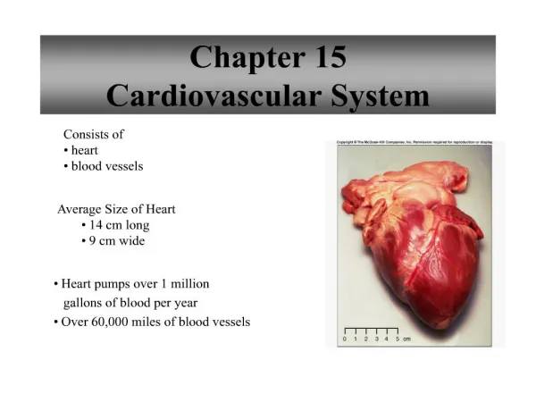
Introduction • Cardiovascular system = the heart and its vessels • The heart pumps 7,000 L of blood per day • Contacts 2.5 billion times in a lifetime
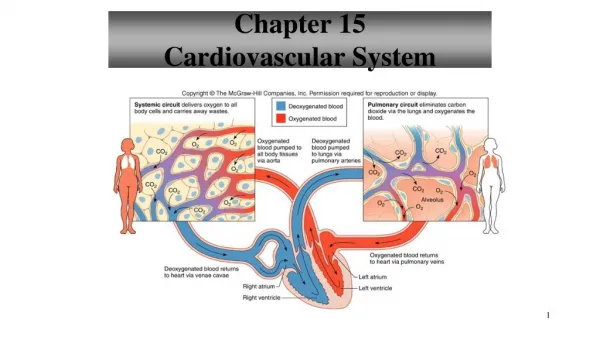
Introduction • Pulmonary circuit = sends deoxygenated blood to the lung where it picks up oxygen and unloads carbon dioxide • Systemic circuit = sends oxygenated blood and nutrients to all body cells and removes wastes
13.2 Structure of the Heart
Size and Location of the Heart • Hollow, cone shaped, muscular pump • Size varies with body size (14 cm by 9 cm) • Base at the top and apex pointing downward
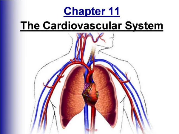
Size and Location of the Heart • Lies within the thoracic cavity and rests on the diaphragm • Within the mediastinum • Bordered laterally by lungs, posteriorly by vertebral column, and anteriorly by sternum
Coverings of the Heart • Pericardium = encloses the heart and proximal ends of the large blood vessels • Has 3 layers • Fibrous pericardium – outer bag of dense connective tissue
Coverings of the Heart • Delicate double layered sac (serous pericardium) • Visceral pericardium – innermost layer covering heart • Parietal pericardium – forms the inner lining of the fibrous pericardium • The two layers are continuous at the base of the heart • Between is the pericardial cavity which is filled with serous fluid (reduces friction)
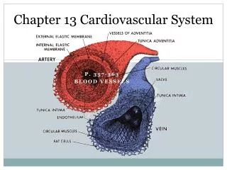
Wall of the Heart • 3 distinct layers • Epicardium – outer layer that corresponds to the visceral pericardium • Myocardium – thick middle layer consisting mostly of cardiac muscle tissue • Endocardium – inner layer consisting of epithelium and connective tissue
Heart Chambers and Valves • Internally divided into 4 hollow chambers • Atria = upper chambers with thin walls that receive blood returning to the heart • Ventricles = lower chambers with thick walls that contract to force blood out of the heart • Septum = solid wall separating left and right sides
Heart Chambers and Valves • The right atrium receives blood from the superior vena cava, the inferior vena cava, and the smaller coronary sinus • Tricuspid valve = permits blood to move from the right atrium to the right ventricle and prevents backflow
Heart Chambers and Valves • Chordaetendineae = strong fibrous strings that attach the cusps of the tricuspid valve • Papillary muscles = small mounds of cardiac muscle that connect chordaetendineae to the walls of the ventricle • Prevents the cusps from swinging back into the atria
Heart Chambers and Valves • The right ventricle must only pump blood to the lungs • Blood exits through the pulmonary valve • Leads to the pulmonary trunk which divides to form the right and left pulmonary arteries
Heart Chambers and Valves • The left atrium receives blood from the lungs through 4 pulmonary veins • Mitral valve (bicuspid valve) = allows blood to pass from the left atrium to the left ventricle and prevents backflow • Also has papillary muscles and chordaetendineae
Heart Chambers and Valves • The left ventricle must pump blood through the aorta to the entire body • Blood exits through the aortic valve
Heart Chambers and Valves • Atrioventricular (A-V) valves • Mitral and tricuspid valves • Between the atria and the ventricles • Semilunar valves • Pulmonary and aortic valves • Name due to the half-moon shapes of the cusps
Skeleton of the Heart • Rings of dense connective tissue surround the pulmonary trunk and aorta • Provide firm attachments for the heart valves and muscle fibers
Path of Blood Through the Heart • Blood that is low in oxygen from the body vena cava and coronary sinus right atrium tricuspid valve right ventricle pulmonary valve pulmonary trunk to the lungs • Blood that is high in oxygen from the lungs pulmonary veins left atrium mitral valve left ventricle aortic valve aorta to the rest of the body
Blood Supply to the Heart • Coronary arteries = first 2 branches of the aorta that supply blood to the tissues of the heart • Blood drains from the heart into the coronary veins which join to form the coronary sinus which empties into the right atrium