Download
1 / 42
420 likes | 453 Vues
Explore the intricacies of cardiology, the medical specialty focusing on heart disorders, and the cardiovascular system that supplies the body with essential blood and oxygen. Learn about the heart's structure, circulation pathways, and the cardiac cycle's vital functions.

E N D
What is Cardiology? • Cardiology: The medical specialty dealing with the diagnosis and treatment of disorders of the heart • Latin: cardium Greek: kardia • Heart’s primary purpose: To supply the body with blood and oxygen it needs to function properly • Pumps blood through a closed circuit of vessels in a continuous loop, carrying oxygen and nutrients from the heart to parts of the body and deoxygenated blood back to the heart to be reoxygenated
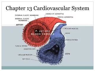
The Cardiovascular System • Blood vessels—A network of interconnecting arteries, arterioles, capillaries, venules, and veins—provide the pathway through which blood is transported between the heart and body cells. Functions: • Arteries and arterioles – distribute • Capillaries – exchange • Veins and venules– collect
Arteries: Carry blood away from the heart to supply organs and tissues with oxygen and nutrients • Arterioles: Smaller arteries which adjust their diameter to increase or decrease blood flow to a particular body tissue • Capillaries: Allow oxygen and nutrients to pass from the blood in the arterioles into tissues and allow waste products to pass from tissues into the blood. • Venules: Small vessels that gather blood from the capillaries • Veins: The larger vessels that carry deoxygenated blood back to the heart
Anatomy of The Heart • The heart is composed almost entirely of muscle • Two Different Pumps: • Pumps deoxygenated blood into the lungs to gather oxygen • Pumps oxygenated blood from the lungs throughout the body
Pericardium • Surrounds the heart like a sac Layers: • Epicardium – outer layer • Myocardium – middle layer • Endocardium – inner layer
Chambers: Sections through which blood is pumped • Right atrium (upper chamber) • Left atrium (upper chamber) • Right ventricle (lower chamber) • Left ventricle (lower chamber)
Valves: Open and close to maintain blood flow through the heart • Tricuspid valve – between right atrium and right ventricle • Mitral valve – between left atrium and left ventricle • Pulmonary valve – between the right ventricle and pulmonary artery • Aortic valve – between the left ventricle and the aorta • Leaflets: Prevent backflow of blood during heartbeat
Coronary arteries: A network of blood vessels that supply oxygen- and nutrient-rich blood directly to the heart’s muscle tissue • Aorta: The main arterial line • Right coronary artery (RCA) • Left coronary artery (LCA) • Posterior descending artery (PDA): Main branch of the RCA • Left main coronary – branches off LCA • Left anterior descending artery (LAD) • Left circumflex artery (LCA) • Two diagonal branches (D1 and D2) • Two obtuse marginal branches (OM1 and OM2)
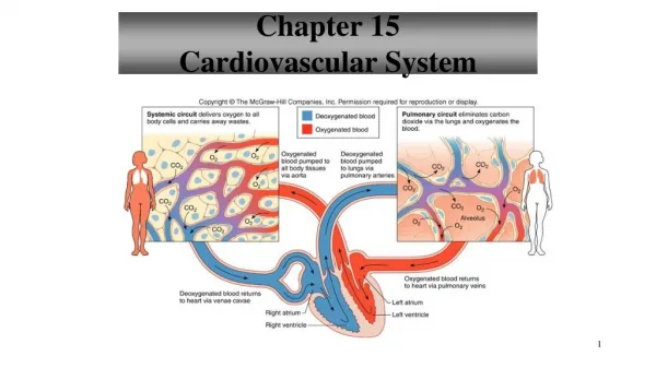
Cardiovascular Circulation • There are two major pathways of the vascular circulation of blood in the body: pulmonary and systemic • Pulmonary circulation is the part of vascular circulation that begins at the right ventricle of the heart • Systemic circulation is the part that begins at the left ventricle
Pulmonary circulation: The right ventricle contracts and pumps blood into the main pulmonary artery, branches off into right and left pulmonary arteries, which deliver deoxygenated blood to the corresponding lung. • In the lungs, each artery branches into smaller arteries and arterioles, and then to capillaries. Gas exchange of oxygen and carbon dioxide takes place between the pulmonary capillaries and the alveoli of the lungs. The oxygenated blood is then returned to the left atrium. This oxygenated blood then travels through the systemic circulation to the rest of the body and the pulmonary circulation begins again.
Systemic circulation: The left ventricle pumps blood into the aorta, which branches off into other arteries that take oxygenated blood into arterioles and capillary networks throughout the body. • After exchange of oxygenated blood and waste products, the deoxygenated blood is sent back to the heart via the superior vena cava and the inferior vena cava. Blood in the right atrium empties in to the right ventricle, and the process starts again.
The Cardiac Cycle • Cardiac Conduction: The name given to the electrical conduction system that controls the heart rate • Provides the electric stimulus necessary for heart to beat • Cardiac Cycle: The sequence of events in one heartbeat • The right and left atria accept blood returning to the heart from the body • The two ventricles push blood out of the heart to be circulated throughout the body • Contraction phase: Systole • Relaxation phase: Diastole
Process: • Electrical stimulus from the sinoatrial (SA) node, “the heart’s natural pacemaker” • To the atrioventricular (AV) node – acts as a bridge between the atria and ventricles in regulating the electrical impulse from the SA node • To the bundle of His – fiber bundles that carry impulse throughout the ventricles • To the left and right bundle branches (pathways) • To the Purkinje fibers – microscopic muscle branches which trigger the ventricles to contract and push blood out into the lungs and body • Cycle begins again
Heart Sounds • Heart sounds: Vibrations in the tissues and blood caused by closure of the heart valves • Lub-dub: Healthy sound, called first heart sound (S1, the lub) and second heart sound (S2, the dub) • Murmurs: Abnormal heart sounds • Graded based on degree to which they are audible • Six-point grading system, with 1 being barely detectable and 6 being very loud • Expressed with a slash between murmur grade and scale used (e.g., grade 1/6 murmur)
A rub is an abnormal sound that is caused by the friction between the beating heart and the pericardium and resembles the sound of squeaky leather and often is described as grating, scratching, or rasping. • A gallop is a tripling or quadrupling of heart sounds that includes three or four sounds that resemble the cantering of a horse. • A click, or systolic click, is a short, high-pitched sound heard when a valve is not functioning properly and may be indicative of valvular disease. • A thrill is a high-frequency vibration felt on the chest wall over the heart, which may be indicative of a structural defect of the heart.
Heart Rates and Rhythms • Normal heart rate is about 60-80 beats per minute (pulse), called sinus rhythm • Arrhythmia: Lack of a normal heart rhythm (or dysrhythmia)
Abnormal heart rates and rhythms • Bradycardia: Slow heartbeat, less than 60 bpm • Tachycardia: Fast heart rate, greater than 100 bpm • Atrial flutter: Atrial rhythm is regular, but the rate isabnormally fast • Fibrillation: Uncoordinated, irregular contraction of theheart muscle • Atrial fibrillation • Ventricular fibrillation
Heart block: Impaired conduction of the heart’s electrical impulses • Paroxysmal atrial tachycardia: A rapid heart rate that starts and stops suddenly and unpredictably • Premature atrial contraction: An extra heartbeat thatoriginates from the atria before it should
Blood Pressure • Measures the force of blood against artery walls • Sphygmomanometer: Measures blood pressure • Indicated in terms of millimeters of mercury (mmHg) and expressed as a fraction—e.g., 120/80 • Systolic pressure (top number): Maximum pressure on the arteries as the heart contracts and pumps blood into the arteries • Diastolic pressure (bottom number): Minimum pressure as the heart relaxes following a contraction
Hypotension: A blood pressure reading of 90/60 mmHg or lower • Orthostatic hypotension: The sudden temporary decrease in systolic blood pressure that occurs when a person changes position, resulting in a feeling of lightheadedness
Common Cardiac Diseases and Treatments • Signs of heart problems • Cyanosis: bluish tinge • Pallor: paleness • Edema: swelling of tissues • Diaphoresis: excessive sweating • Angina (angina pectoris): severe chest pain
Hypertension (high blood pressure): A condition in which the pressure of the blood in the arteries is too high, possibly damaging the heart and blood vessels • Primary hypertension: No identifiable cause • Secondary hypertension: Another known cause (disease) Treatments: • Diuretics: Eliminate extra sodium and fluid from the body • Beta-blockers: Decrease heart rate and the amount of blood the heart pumps out with each beat • ACE inhibitors: Inhibit the formation of angiotensin II, which narrows blood vessels • Calcium-channel blockers: Decrease the force of the heart’s pumping action • ARBs: Reduce pressure like ACE inhibitors but without some of the side effects
Coronary Artery Disease (CAD): Narrowing arteries hinder blood supply to heart • Also called cardiac ischemia: From ischi, to hold back and –emia, blood • Cause: Plaques (atherosclerosis) build up in arteries, causing arteries to narrow and harden, reducing blood flow, causing stenosis (narrowing), anginapectoris (severe chest pain), and dyspnea (shortness of breath)
Complications: • Heart failure: The heart does not pump the way it should • Congestive heart failure (CHF): Heart’s weak pumping action causes fluid buildup (congestion) in the lungs and other body tissues • Orthopnea: Breathing difficulties while lying down • Paroxysmal nocturnal dyspnea (PND): A sudden onset of breathing difficulty occurring at night. Term breakdown: • Paroxysmal – sudden onset of a symptom • Nocturnal – pertaining to hours of night • Dyspnea – difficulty breathing
Treatments for CAD: • Nitrates (nitroglycerin): Relieve angina by dilating blood vessels • Antihypertensive medications • Surgical interventions: • Percutaneous transluminal coronary angioplasty (PTCA) • Opens narrowed arteries using a catheter, a thin flexible tube with a tiny balloon attached • The balloon inflates in the artery, flattening the plaque against the artery wall and enabling better blood flow • Stent is implanted at the site to keep the artery from collapsing
Treatments for CAD (Continued) • Surgical interventions: • Coronary artery bypass graft (CABG): Restores circulation when occluded, or blocked, coronary arteries prevent normal blood flow to the heart • Bypasses around the blockages are performed using different vessels as grafts • Greater saphenous vein in leg and thigh most common, called a saphenous vein graft • The new, healthy graft carries blood around the blockage
Cardiomyopathy: Impairment in structure and function of the myocardium • Dilated cardiomyopathy: Enlargement of the heart chambers • Hypertrophic cardiomyopathy: Overgrowth of heart muscle • Restrictive cardiomyopathy: Ventricles stiffen and do not fill normally Treatment: • Drug therapy (ACE inhibitors, anticoagulants, diuretics)
Valvular Heart Disease: The heart’s ability to pump blood is impeded due to valve malfunction • Blood leaks back through valves that should be closed (regurgitation) • Mitral valve prolapse: The heart’s mitral valve bulges slightly back into the left atrium when it closes, causing regurgitation into the atrium • Treatment: Not usually required but if severe, surgical treatment to repair valve is performed • Patients may be prescribed antibiotics before dental or other procedures to prevent bacterialendocarditis, a bacterial infection which could lead to deformity and destruction of valve leaflets
Pericarditis: An inflammation of the pericardium that surrounds the heart • The amount of fluid between the inner and outer layers of the heart increases, compressing the heart and interfering with its function • Pericardial effusion: Fluid accumulates in the pericardial sac, putting pressure on the heart and causing it not to fill adequately or pump properly • Cardiac tamponade: Less blood leaves the heart, causing dramatic drop in blood pressure. Potentially fatal if not treated immediately!
Causes of pericarditis: • Bacterial or fungal infection • AIDS, cancer, or tuberculosis • After a heart attack • Dressler syndrome: Delayed onset of pericarditis • Treatments: • Mild cases heal on their own • Pericardiocentesis: Drainage of excess fluid (also called pericardial window) • Pericardiectomy: Portion of diseased pericardium is removed
Congenital Heart Disorders • Atrial septal defect (ASD): A “hole in the heart,” or in atrial septum • Treatment: Hole usually closes on its own • Ventricular septal defect (VSD): A hole in the ventricular septum • Treatment: Hole usually closes on its own
Patent Ductus Arteriosus (PDA) • Ductus arteriosus remains open (patent) after birth • Treatment: Condition will resolve alone or with corrective surgery • Transposition of the Great Vessels • Location of aorta and pulmonary artery (great vessels) is switched • Treatment: Arterial switch operation
Tetralogy of Fallot • Too little oxygen in the blood, leading to cyanosis • Combination of four defects: • VSD • Pulmonary stenosis • Displaced aorta • Right ventricular hypertrophy • Treatment: Surgery to increase blood flow and correct defects
Diagnostic Studies and Procedures • Blood Tests • Cardiac risk factor testing: Evaluate risk of heart disease or stroke • C-reactive protein (CRP) • Homocysteine • Lipoprotein (a) or Lp(a) (dictated as “L, P, little A”) • Cholesterol particle test
Lipid profile (total cholesterol, HDL and LDL) • Blood sugar (glucose) • B-type natriuretic peptide (BNP) • Cardiac enzyme studies
Electrocardiogram (EKG) • Analyzes electrical activity of the heart • During the test, electrodes called leads are placed on patient’s arms, legs, and chest wall • The impulses detected by the leads are produce graphic tracing of waveforms on paper strip • Deviation in shape or interval of waveforms indicates possible problem
Echocardiogram • Uses ultrasound to examine heart anatomy • Sound waves “echo” off cardiac structures • Provides image of beating heart on a computer screen
Cardiac Stress Test: Evaluates the heart when heart is working • Measured using Bruce protocol • Expressed in metabolic equivalents (METS) • Nuclear scan thallium • Camera records thallium received by the heart • Shows problems with blood flow and pumping action
Cardiac Catheterization and Angiography • Diagnose and treat heart disorders other than those involving coronary arteries • Radiopaque dye is inserted into coronary arteries to view images of the blood vessels as the heart pumps
Multiple gated acquisition (MUGA) scan • Determines if the left and right ventricles are functioning properly • Diagnoses abnormalities in the heart wall
Insight The Heart-Brain