Download
1 / 81
810 likes | 978 Vues
Cardiovascular System Chapters 19 Blood. Anatomy and Physiology Liberty Senior High Mr. Knowles. What’s the purpose of the cardiovascular system? Do all organisms have one?. Let’s model!. Why we need a cardiovascular system!.

E N D
Cardiovascular SystemChapters 19 Blood Anatomy and Physiology Liberty Senior High Mr. Knowles
What’s the purpose of the cardiovascular system?Do all organisms have one? Let’s model!
Why we need a cardiovascular system! • Human embryos before 3 weeks are so small, materials are transported by simple diffusion. • At third week (few mms in length), heart begins beating- first system to function. • Supplies nutrients to all 75 trillion cells in the body.
What is the cardiovascular system? Three parts: • Blood – a circulating fluid. (Chapter 19). • Heart – a pump. (Chapter 20). • Blood vessels – the conducting pipes (Chapter 21)
Cardiovascular Lymphatic Systems • Fluid leaves the vessel and enters the tissues- interstitial fluid. • Eventually returns to the vessels. • Lymphatic system has its own vessels. • Used to transport antibodies, white blood cells, and monitor for infection and cancer. • Cardiovascular + Lymphatic = Circulatory System.
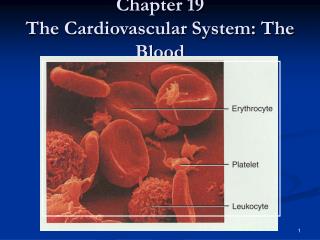
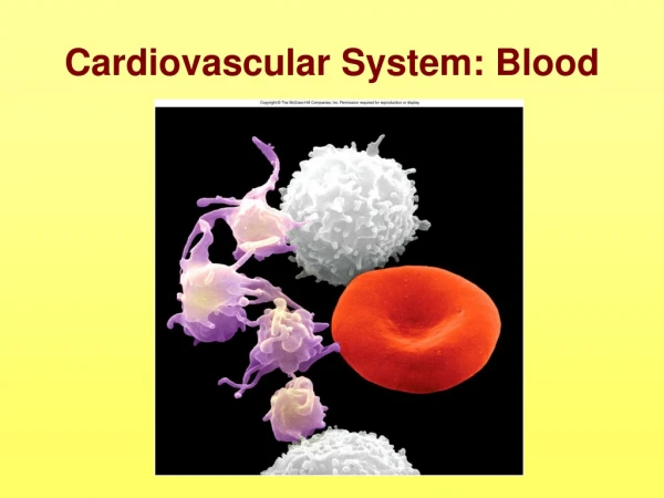
What is blood? • Specialized connective tissue with cells in a fluid matrix.
Functions of the Blood • Transport dissolved gases, nutrients, hormones, and metabolic wastes. • Regulation of the pH and electrolytes of interstitial fluid. Neutralizes the acids created by metabolism (lactic acid). • Restricts fluid losses through damaged vessels or at injury sites- blood clots.
Functions of the Blood • Defense against toxins and pathogens- transports white blood cells that migrate into tissue to fight infection and remove debris. Also, deliver antibodies. • Stabilize body temperature- absorbs heat from active muscles and distributes to other tissues. Also brings heat to the surface of the skin to lose heat.
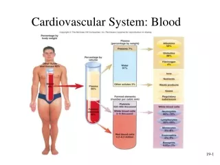
Composition of Blood • It is a fluid connective tissue with an extracellular matrix- plasma + formed elements (cells and cell fragments) = whole blood. • Plasma + Formed Elements = Whole Blood.
Whole Blood After Centrifugation Plasma White Blood Cells “Buffy Coat” Red Blood Cells
Whole Blood 37-54% 46-63% Centrifuge and Separate Formed Elements Plasma
Plasma 1 % Electrolytes and other Solutes 7 % Plasma Proteins 92 % Water
Plasma- The Fluid of Life! • Plasma = Plasma Proteins + a Ground Substance (Serum). • Plasma Proteins: Albumin- transport fatty acids, maintain isotonic solution. Globulin- immunoglobulin (antibodies). Fibrinogen- form blood clots; becomes fibrin- an insoluble protein.
Plasma Globulin Serum Albumin Fibrinogen
Plasma- The Fluid of Life! • Plasma that has been allowed to clot will lose its fibrin and other salts like Ca+2. • Plasma without its fibrin – Serum.
Formed Elements • Formed Elements = Blood Cells + Fragments suspended in the plasma. • Erythrocytes (Red Blood Cells) – most abundant (99.9% of all cells); transport of oxygen and carbon dioxide. • Leukocytes (White Blood Cells) – body’s defense cells. (0.1% of cells). • Thrombocytes (Platelets) – small, membrane- bound packets of cytoplasm that contain enzymes for blood clot formation.
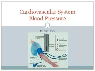
Collecting and Analysis of Blood • Blood usually collected at a vein-venipuncture. • Venipuncture- veins are easy to locate, walls of vein are thinner, pressure is lower heals easier. • Peripheral capillaries- tip of finger, earlobe; oozing small drop for blood smear. • Arterial Puncture- check for efficiency of gas exchange.
Properties of Blood • Temperature- 38° C or 100.4° F. • Viscosity- has a great deal of dissolved proteins in plasma more viscous than water. • pH – 7.35-7.45; slightly alkaline.
Erythrocytes (RBCs) • “erythros”- red; “cyte”- cell. • RBCs are the most abundant blood cell (99.9%). 25 trillion in average adult. Takes ~ 1 min. to travel circuit. • Hematocrit- percentage of formed elements in a sample of whole blood. # of cells / microliter of whole blood. • Has a red pigment-hemoglobin- gives whole blood its color.
RBCs Structure and Function • Highly specialized cell to transport gases. • Cell structure is a biconcave disc.
RBCs Structure and Function • Shape provides the RBC with a large surface area. • Exchange of O2 with the surrounding plasma must be quick; larger surface area faster the exchange. • Total surface of all RBCs is 3800 m2 compared to 1.9 m2 of the whole human body.
RBCs Structure and Function • Biconcave shape allows them to form stacks (dinner plates) – rouleaux inside narrow blood vessels. • Rouleaux permit the cells to pass through blood vessels without bumping along the walls. • Do not form logjams or clogs in the narrow capillary.
RBCs Structure and Function • Biconcave shape allows the RBCs to bend and flex when entering capillaries. • May pass through capillaries ½ the RBC’s diameter.
RBC’s are Highly Specialized Cells • Have lost all organelles- lack nuclei, mitochondria, and ribosomes. • Lost these structures to allow more space for hemoglobin and oxygen transport. • Downside: RBCs unable to divide or repair themselves. Made in bone marrow. • Short lifespan- 120 days and then must be broken down.
Hemoglobin (Hb) • Accounts for 95% of proteins inside the RBC. • 280 million Hbs in each RBC. • Hb binds to and transports O2 and CO2.
Hb Molecule • Each Hb molecule = four protein chains = 2 alpha chains + 2 beta chains of polypeptides. • Each chain is a globular subunit and has a heme group. • Heme – a porphyrin which is a ring compound with an iron in the center. • Iron has a + charge and can bind to O2 (negative).
Hb Molecule • When hemoglobin binds to O2 – it becomes oxyhemoglobin. • Very weak interaction; easy to separate. • Fetus uses a fetal hemoglobin- more readily binds to O2 for more efficient uptake from mother’s RBCs.
Hb Molecule • Alpha and Beta chains bind to CO2 at other sites and transport to lungs. • If hematocrit is low or the amount of Hb in RBCs is low than normal activity cannot be sustained in tissue- anemia.
Sickle Cell Anemia • Mutations in the beta chains of the Hb molecule. • When the blood contains abundant O2, the Hb and RBCs are normal. • But when the defective Hb loses its O2, neighboring Hb molecules interact and change the shape of the cell- curved and stiff. • Cannot form rouleaux and may form clots.
Sickle Cell Mutation Sickle Cell Mutation
Leukocytes (WBCs) • General Properties: 1. Help defend against pathogens, toxins, and damaged cells. 2. They have nuclei and other organelles. 3. Are made in bone marrow, thymus, spleen, and other lymphatic tissue.
Two Major Groups of WBCs • Granulocytes- WBCs with darkly-staining vesicles and lysosomes inside. a. Neutrophils b. Eosinophils c. Basophils