Higher Order Protein Structures
Higher Order Protein Structures. Lecture 6, Medical Biochemistry. Lecture 6 Outline. Molecular forces involved in protein structure Protein folding Structure and biosynthesis of collagen. Protein Native State and Denaturation. A protein folded into its tertiary

Higher Order Protein Structures
E N D
Presentation Transcript
Higher Order Protein Structures Lecture 6, Medical Biochemistry
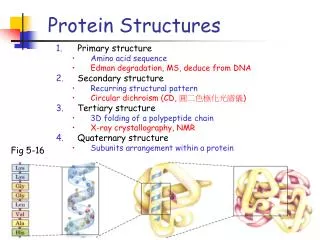
Lecture 6 Outline • Molecular forces involved in protein structure • Protein folding • Structure and biosynthesis of collagen
Protein Native State and Denaturation A protein folded into its tertiary structure under optimal conditions is said to be in its “native state,” which corresponds to its most thermo- dynamically favorable arrangement of atoms. Proteins can be denatured (loss of their secondary and higher structures) by changes in temperature, pH, ionic strength, urea or detergents
Proposed Folding Pathway Accessory proteins?
Compare: Bond kJ/m C-C = 350 C-H = 410 O-H = 460
Non-covalent Molecular Forces in Protein Structure and Folding • Hydrophobic interactions: interactions between hydrophobic amino acids is likely the largest noncovalent force responsible for most protein folding. Recall that water will tend to form a solvation shell around free hydrophobic compounds, which in thermodynamic terms, is a decrease in entropy (and thus not favored). This drives formation of hydrophobic regions of protein chains to come together, the water shell is disrupted and entropy increases.
Non-covalent Forces (cont) • For hydrophobic interactions, calculations have shown that 1/3 loss of water of solvations occurs with formation of secondary structure; an additional 1/3 water of solvation is lost in the formation of tertiary structure. This is a large source of the energy that drives protein folding. • Hydrogen Bonds - these have been discussed for helix and sheet formation. Distance between the donor and acceptor atoms are the most important determinants for bond formation, 2.7-3.1 angstroms are optimal (covalent bond is 1.0-1.6 angstroms)
Non-covalent Forces (cont) • Electrostatic, or charge-charge interactions. Predominantly found on the exterior surfaces of proteins, interacting with the water solvent. The strength of the ionic forces are largely dampened by the high dielectric constant of the surrounding water, such that proximity to other charged groups is likely the strongest attractive force for charge-charge interactions
Non-covalent Forces (cont) • Van der Waals interactions: Occur when molecules or atoms which do not have covalent bonds between them come so close together that their outer electron orbitals begin to overlap. This can lead to changes in the overall distribution of the electronic charges to create a weak attractive force. Contact distance ranges from 2.8 to 4.1 angstroms, so forces are weak. However, in a folded protein, thousands of individual interactions occur, thus providing a significant cumulative stabilizing force.
Accessory Proteins for Folding • In a test tube, protein renaturation of an unfolded protein can take minutes, days or never occur. Protein folding in the cell occurs during protein translation (synthesis) and generally takes only a few minutes for formation of the native conformation. This is primarily due to the inherent properties of the protein, and is assisted by cellular accessory proteins, examples of which include:
Accessory Proteins (cont) • Protein disulfide isomerase - catalyzes formation of disulfide bonds • Peptidyl Prolyl Cis-Trans Isomerases - this class of enzymes, also called rotamases, facilitates the conversion of Pro residues to cis-conformations (originally made in a trans conformation)
Accessory Proteins (cont) • Chaperones - a large group of proteins, also termed heat shock proteins and chaperonins, their precise regulation and mechanisms of action remain largely undefined. During the folding process, they function to prevent unfavorable protein interactions with other potentially complementary surfaces (like other proteins, carbohydrates, lipids, nucleic acids, etc.) Many of these proteins are ATPases (use hydrolysis of ATP as an energy source).
Fibrous Proteins - Examples and Characteristics • Highly elongated proteins whose secondary structures are the dominant structural motif • Found in skin, muscle, tendon and bone and have connective, protective and/or supportive functions • EXAMPLES: keratins (hair, nails, horn,feathers) elastin (tendons); spider silk fibroin; collagens
COLLAGEN: Properties • Most abundant protein in mammals • example of a fibrous protein (in general, these protein have repetitive secondary structures, are poorly soluble, and cannot be crystallized; other fibrous proteins include elastin in tendons & keratins in hair and nails) • collagen composed of approximately 33% glycine, 21% proline or hydroxyproline, and 11% alanine
COLLAGEN: Properties (cont) • Tropocollagen (the precursor form) is composed of three polypeptide chains of about 1000 amino acids each, wrapped in a triple helix, rope-like coil • Every third residue in each polypeptide in this triple helix is a glycine. The glycines from each strand interact with each other in the central, shared interior of the helix • Proline frequently follows glycine, and the third residue can be any other amino acid (X) found in collagen; a Gly-Pro-X motif
COLLAGEN: Properties (cont) • Extensive post-translational modification of prolines and lysines occurs • Cross-linked fibrils of collagen form after secretion and processing of tropocollagen • Defects in enzymes involved in the collagen biosynthetic pathway are responsible for many diseases.
Formation of collagen cross-links: Mediated by lysine modifications and subsequent lysine-allysine or allysine-allysine covalent bond formation
Post-translational processing of collagen
Marfan syndrome is caused by mutations in the fibrillin gene; Fibrillin is a large fibrous protein component of extracullar microfibrils, frequently associated with elastin