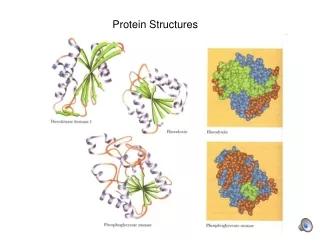
Protein structures
Explore the importance of protein structure in biological functions, the basics of proteins, various measurements for protein structure, and the different levels of protein structure. Learn about the Protein Data Bank and the basics of primary, secondary, tertiary, and quaternary structures.

Protein structures
E N D
Presentation Transcript
Protein Structure • Why protein structure? • The basics of protein • Basic measurements for protein structure • Levels of protein structure • Prediction of protein structure from sequence • Finding similarities between protein structures • Classification of protein structures
Why protein structure? • In the factory of living cells, proteins are the workers, performing a variety of biological tasks. • Each protein has a particular 3-D structure that determines its function. • Protein structure is more conserved than protein sequence, and more closely related to function.
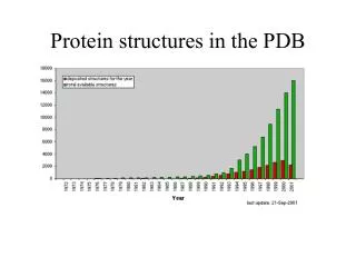
Structural information • Protein Data Bank: maintained by the Research Collaboratory of Structural Bioinformatics(RCSB) • http://www.rcsb.org/pdb/ • > 42752 protein structures as of April 10 • including structures of Protein/Nucleic Acid Complexes, Nucleic Acids, Carbohydrates • Most structures are determined by X-ray crystallography. Other methods are NMR and electron microscopy(EM). Theoretically predicted structures were removed from PDB a few years ago.
PDB Growth Red: Total Blue: Yearly
The basics of proteins • Proteins are linear heteropolymers: one or more polypeptide chains • Building blocks: 20 types of amino acids. • Range from a few 10s-1000s • Three-dimensional shapes (“fold”) adopted vary enormously.
Basic Measurements for protein structure • Bond lengths • Bond angles • Dihedral (torsion) angles
Bond Length • The distance between bonded atoms is constant • Depends on the “type” of the bond • Varies from 1.0 Å(C-H) to 1.5 Å(C-C) • BOND LENGTH IS A FUNCTION OF THE POSITIONS OF TWO ATOMS.
Bond Angles • All bond angles are determined by chemical makeup of the atoms involved, and are constant. • Depends on the type of atom, and number of electrons available for bonding. • Ranges from 100° to 180° • BOND ANGLES IS A FUNCTION OF THE POSITION OF THREE ATOMS.
Dihedral Angles • These are usually variable • Range from 0-360° in molecules • Most famous are , , and • DIHEDRAL ANGLES ARE A FUNCTION OF THE POSITION OF FOUR ATOMS.
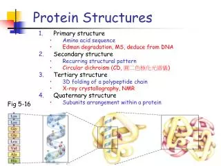
Levels of protein structure • Primary structure • Secondary structure • Tertiary structure • Quaternary structure
Primary structure • This is simply the amino acid sequences of polypeptides chains (proteins).
Secondary structure • Local organization of protein backbone: -helix, -strand (groups of -strands assemble into -sheet), turn and interconnecting loop. an a-helix various representations and orientations of a two stranded b-sheet.
The -helix • One of the most closely packed arrangement of residues. • Turn: 3.6 residues • Pitch: 5.4 Å/turn
The -sheet • Backbone almost fully extended, loosely packed arrangement of residues.
Catechol O-Methyltransferase -Sheet (parallel) All strands run in the same direction
Urate oxidase -Sheet (antiparallel) All strands run in the opposite direction, more stable
Loops and Turns Loops: often contain hydrophilic residue on the surface of proteins Turns: loops with less than 5 residues and often contain G, P
Tertiary structure • Description of the type and location of SSEs is a chain’s secondary structure. • Three-dimensional coordinates of the atoms of a chain is its tertiary structure. • Quaternary structure: describes the spatial packing of several folded polypeptides
Tertiary structure • Packing the secondary structure elements into a compact spatial unit • “Fold” or domain– this is the level to which structure prediction is currently possible.
Quaternary structure • Assembly of homo or heteromeric protein chains. • Usually the functional unit of a protein, especially for enzymes
Primary and secondary structure are ONE-dimensional; Tertiary and quaternary structure are THREE-dimensional. • “structure” usually refers to 3-D structure of protein.
PDB Files: the “header” HEADER OXIDOREDUCTASE(SUPEROXIDE ACCEPTOR) 13-JUL-94 COMPND MANGANESE SUPEROXIDE DISMUTASE (E.C.1.15.1.1) COMPLEXED COMPND 2 WITH AZIDE OURCE (THERMUS THERMOPHILUS, HB8) AUTHOR M.S.LAH,M.DIXON,K.A.PATTRIDGE,W.C.STALLINGS,J.A.FEE, AUTHOR 2 M.L.LUDWIG REVDAT 2 15-MAY-95 REVDAT 1 15-OCT-94 JRNL AUTH M.S.LAH,M.DIXON,K.A.PATTRIDGE,W.C.STALLINGS, JRNL AUTH 2 J.A.FEE,M.L.LUDWIG JRNL TITL STRUCTURE-FUNCTION IN E. COLI IRON SUPEROXIDE JRNL TITL 2 DISMUTASE: COMPARISONS WITH THE MANGANESE ENZYME JRNL TITL 3 FROM T. THERMOPHILUS JRNL REF TO BE PUBLISHED REMARK 1 AUTH M.L.LUDWIG,A.L.METZGER,K.A.PATTRIDGE,W.C.STALLINGS REMARK 1 TITL MANGANESE SUPEROXIDE DISMUTASE FROM THERMUS REMARK 1 TITL 2 THERMOPHILUS. A STRUCTURAL MODEL REFINED AT 1.8 REMARK 1 TITL 3 ANGSTROMS RESOLUTION REMARK 1 REF J.MOL.BIOL. V. 219 335 1991 REMARK 1 REFN ASTM JMOBAK UK ISSN 0022-2836 REMARK 1 REFERENCE 2 REMARK 1 AUTH W.C.STALLINGS,C.BULL,J.A.FEE,M.S.LAH,M.L.LUDWIG REMARK 1 TITL IRON AND MANGANESE SUPEROXIDE DISMUTASES: REMARK 1 TITL 2 CATALYTIC INFERENCES FROM THE STRUCTURES
PDB Files: the coordinates Atom & Residue XYZ Coordinates ATOM 1 N PRO A 1 10.846 26.225 -13.938 1.00 30.15 1MNG 192 ATOM 2 CA PRO A 1 12.063 25.940 -14.715 1.00 28.55 1MNG 193 ATOM 3 C PRO A 1 12.061 26.809 -15.946 1.00 26.55 1MNG 194 ATOM 4 O PRO A 1 11.151 27.612 -16.176 1.00 26.17 1MNG 195 ATOM 5 CB PRO A 1 12.010 24.474 -15.162 1.00 30.21 1MNG 196 ATOM 6 CG PRO A 1 11.044 23.902 -14.231 1.00 31.38 1MNG 197 ATOM 7 CD PRO A 1 9.997 25.028 -14.008 1.00 31.86 1MNG 198 ATOM 8 N TYR A 2 13.050 26.576 -16.777 1.00 23.36 1MNG 199 ATOM 9 CA TYR A 2 13.197 27.328 -17.983 1.00 22.11 1MNG 200 ATOM 10 C TYR A 2 12.083 27.050 -19.032 1.00 21.02 1MNG 201 ATOM 11 O TYR A 2 11.733 25.895 -19.264 1.00 21.68 1MNG 202 ATOM 12 CB TYR A 2 14.579 26.999 -18.523 1.00 20.16 1MNG 203 ATOM 13 CG TYR A 2 14.905 27.662 -19.832 1.00 19.42 1MNG 204 ATOM 14 CD1 TYR A 2 14.516 27.092 -21.038 1.00 18.28 1MNG 205 ATOM 15 CD2 TYR A 2 15.610 28.864 -19.875 1.00 19.69 1MNG 206 ATOM 16 CE1 TYR A 2 14.813 27.696 -22.233 1.00 19.13 1MNG 207 ATOM 17 CE2 TYR A 2 15.924 29.465 -21.070 1.00 19.25 1MNG 208 ATOM 18 CZ TYR A 2 15.515 28.863 -22.251 1.00 19.25 1MNG 209 ATOM 19 OH TYR A 2 15.857 29.417 -23.448 1.00 21.67 1MNG 210 ATOM 20 N PRO A 3 11.583 28.094 -19.731 1.00 19.90 1MNG 211 ATOM 21 CA PRO A 3 11.912 29.520 -19.665 1.00 18.36 1MNG 212
Motifs Helix-loop-helix Four helix bundle Coiled coil
Secondary structure prediction • Given a protein sequence (primary structure) GHWIATRGQLIREAYEDYRHFSSECPFIP • Predict its secondary structure content (C=coils H=Alpha Helix E=Beta Strands) CEEEEECHHHHHHHHHHHCCCHHCCCCCC
Why Secondary Structure Prediction? • Easier problem than 3D structure prediction (more than 40 years of history). • Accurate secondary structure prediction can be an important information for the tertiary structure prediction • Improving sequence alignment accuracy • Protein function prediction • Protein classification • Predicting structural change
Prediction Methods • Statistical methods • Chou-Fasman method, GOR I-IV • Nearest neighbors • NNSSP, SSPAL • Neural network • PHD, Psi-Pred, J-Pred • Support vector machine
Assumptions • The entire information for forming secondary structure is contained in the primary sequence. • Side groups of residues will determine structure. • Examining windows of 13 - 17 residues is sufficient to predict structure.
Chou-Fasman method • Compute parameters for amino acids • Preference to be in • alpha helix: P(a) • beta sheet: P(b) • Turn: P(turn) • Frequencies with which the amino acid is in the 1st, 2nd, 3rd, and 4th position of a turn: f(i), f(i+1), f(i+2), f(i+3). • Use a sliding window
SSE prediction • Alpha-helix prediction • Find all regions where 4 of the 6 amino acids in window have P(a) > 100. • Extend the region in both directions unless 4 consecutive residues have P(a) < 100. • If Σ P(a) > Σ P(b) then the region is predicted to be alpha-helix. • Beta-sheet prediction is analogous. • Turn prediction • Compute P(t) = f(i) + f(i+1) + f(i+2) + f(i+3) for 4 consecutive residues. • Predict a turn if • P(t) > 0.000075 (check) • The average P(turn) > 100 • Σ P(turn) > Σ P(a) and Σ P(turn) > Σ P(b)
GOR method • Use a sliding window of 17 residues • Compute the frequencies with which each amino acid occupies the 17 positions in helix, sheet, and turn. • Use this to predict the SSE probability of each residue.
Performance of SSE prediction Q3 and SOV are standards for computing errors A Simple and Fast Secondary Structure Prediction Method using Hidden Neural Networks Kuang Lin, Victor A. Simossis, Willam R. Taylor, Jaap Heringa, Bioinformatics Advance Access published September 17, 2004
Relevance of Protein Structurein the Post-Genome Era structure medicine sequence function
Structure-Function Relationship Certain level of function can be found without structure. But a structure is a key to understand the detailed mechanism. A predicted structure is a powerful tool for function inference. Trp repressor as a function switch
Structure-Based Drug Design Structure-based rational drug design is a major method for drug discovery. HIV protease inhibitor
Experimental techniques for structure determination • X-ray Crystallography • Nuclear Magnetic Resonance spectroscopy (NMR) • Electron Microscopy/Diffraction • Free electron lasers ?