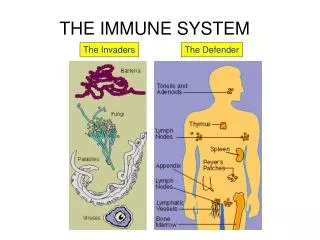
The Immune System
The Immune System. Body Defenses. Reconnaissance, Recognition, and Response Must defend from the many dangerous pathogens it may encounter in the environment Detect invader/foreign cells Communicate alarm & recruit immune cells Suppress or destroy invader

The Immune System
E N D
Presentation Transcript
Body Defenses • Reconnaissance, Recognition, and Response • Must defend from the many dangerous pathogens it may encounter in the environment • Detect invader/foreign cells • Communicate alarm & recruit immune cells • Suppress or destroy invader • Two major kinds of defense have evolved that counter these threats • Innate immunity and acquired immunity
3m Innate immunity • Innate immunity provides broad defenses against infection • Present before any exposure to pathogens and is effective from the time of birth • Involves nonspecific responses to pathogens • A pathogen that successfully breaks through an animal’s external defenses encounters several innate cellular and chemical mechanisms that impede its attack on the body
Innate Immunity • Non-selective • No lag time – immediate response No previous exposure required • Protects against infections, toxins • Works with specific (acquired) immune response
Acquired (Adaptive) Immune Response • Depends on B and T lymphocytes • Specific immune response directed attack against pathogens (antigen) • Lag time • Previous Antigen exposure required • Protects against pathogens and cancer cells • Types • Antibody-mediated: B cells • Cell-mediated: T cells
Types of Immunity Figure 22.14
INNATE IMMUNITY Rapid responses to a broad range of microbes ACQUIRED IMMUNITY Slower responses to specific microbes External defenses Internal defenses Skin Phagocytic cells Humoral response (antibodies) Mucous membranes Antimicrobial proteins Secretions Inflammatory response Invading microbes (pathogens) Cell-mediated response (cytotoxic lymphocytes) Natural killer cells Body Defenses
Innate Immunity • Physical barriers, secretion, chemical toxins • Phagocytosis - macrophages neutrophils engulf and digest recognized "foreign" cells – molecules • Inflammatory response - localized tissue response to injury producing swelling, redness, heat, pain • Natural Killer cells – special class of lymphocyte-like cells that destroy virus infected cells and cancer cells • Complements system activated proteins that destroy pathogen plasma membranes and enhance phagocytosis, inflamation • Interferon - proteins that non-specifically defend against viral infection
Innate Immunity / External Defenses • Physical barriers prevent entry of microorganisms and viruses • Epidermis • Mucous Membranes • Hair, Cilia • Excretions - lacrimal, saliva • Chemical • Skin acidity between 3 and 5, which is acidic enough to prevent colonization • Sebum toxic to microbes • Lysozymes digests the cell walls of many bacteria 10m • In the trachea, ciliated epithelial cells sweep mucus and any entrapped microbes upward, preventing the microbes from entering the lungs
Innate Immunity - Phagocytosis • Scavenge dead, dying body cells, remove cellular debris • Destroy abnormal (cancerous) • Protect from pathogens & foreign molecules: parasites, bacteria, viruses • Monocyte - macrophage system – free and fixed • Margination – stick to the inner endothelial lining of capillaries of affected tissue • Move by diapedesis – move thru capillary walls • Microphages – Neutrophils and eosinophils • Exhibit chemotaxis
Innate Immunity - Phagocytosis Neutrophils • Fastest response of all WBC to bacteria and parasites • Direct actions against bacteria • Release lysozymes which destroy/digest bacteria • Release defensive proteins that act like antibiotics • Release strong oxidants (bleach-like, strong chemicals ) that destroy bacteria Eosinophils • Leave capillaries to enter tissue fluid • Attack parasitic worms • Phagocytize antibody-antigen complexes
Innate Immunity - Phagocytosis Monocytes • Take longer to get to site of infection, but arrive in larger numbers • Become free (roaming) macrophages, once they leave the capillaries • Destroy microbes and clean up dead tissue following an infection
Pseudopodia surround microbes. 1 Microbes Microbes are engulfed into cell. 2 MACROPHAGE Vacuole containing microbes forms. 3 Vacuole Lysosome containing enzymes Vacuole and lysosome fuse. 4 Toxic compounds and lysosomal enzymes destroy microbes. 5 Microbial debris is released by exocytosis. 6 Phagocytic Cells • Phagocytes attach to their prey via surface receptors and engulf them, forming a vacuole that fuses with a lysosome
Phagocytosis Mechanisms • Chemotaxis • Attraction to certain chemical mediators • Released at the site of damage • Chemotaxins induce phagocytes to injury • Opsonization • Identify (mark) pathogen • Coated with chemical mediators • Most important opsonins • Toll-like receptors (TLR’s) • Phagocytic cells studded with plasma membrane receptor proteins • Bind with pathogen markers • Recognition - Allow phagocytes to “see” and distinguish from self-cells Figure 24-6: Phagocytosis
Inflammatory Response • Inflammation – histamine release from mast cells and other chemicals released from injured cells promote changes in blood vessels • Changes allow more fluid, phagocytes, and antimicrobial proteins to enter tissues • Effects of inflammation include • Mobilization of local, regional, and systemic defenses • Slow the spread of pathogens • Temporary repair of injury
Blood clot Pin Pathogen Macrophage Blood clotting elements Chemical signals Phagocytic cells Phagocytosis Capillary Red blood cell Chemical signals released by activated macrophages and mast cells at the injury site cause nearby capillaries to widen and become more permeable. Fluid, antimicrobial proteins, and clotting elements move from the blood to the site. Clotting begins. Neutrophils and macrophages phagocytose pathogens and cell debris at the site, and the tissue heals. Chemokines released by various kinds of cells attract more phagocytic cells from the blood to the injury site. 1 4 2 3 Inflammatory Response • Macrophages, mast cells release histamine • Localized vasodilation • Capillary permeability - increased gaps in capillaries bring more WBC's & plasma proteins • Swelling, redness, heat and pain are incidental • Injured cells and phagocytes release cytokines (chemical signalss) • Kinins - stimulate complement system (plasma proteins) • Chemotaxins – attract more phagocytes • Clotting factors – walling off invasion
Natural Killer Cells • Patrol the body and attack virus-infected body cells and cancer cells • Recognize cell surface markers on foreign cells • Destroy cells with foreign antigens • Rotation of the Golgi toward the target cell and production of perforins • Release of perforins by exocytosis • Interaction of perforins causing cell lysis
How Natural Killer Cells Kill Cellular Targets Figure 22.11
Antimicrobial Proteins • Proteins function in innate defense by attacking microbes directly or impeding their reproduction • Complement System - About 30 proteins involved in the lysis of invading cells and helps trigger inflammation • Interferons – small proteins provide innate defense against viruses and help activate macrophages
Complement System • System of inactive proteins produced by liver circulating in blood and on cell membranes • Cascade of plasma complement proteins (C) activated by antibodies or antigens causing cascade of chemical reactions • Direct effect is lysis of microorganisms by destroying target cell membranes • Indirect effects include: • Chemotaxis • Opsonization • Inflammation: recruit phagocytes, B & T lymphocytes
Complement Activation Figure 22.12
Innate Cytokines - Interferons • Small antiviral proteins released by lymphocytes, macrophages, virally infected cells • Type I interferons – Alpha and Beta • Induced during many virus infections • IFN- a: Mainly by leukocytes • IFN- b: Mainly by fibroblast cells • Binds to membranes of adjacent, uninfected cells • Triggers production of proteins that interfere with viral replication • Enhances macrophage, natural killer, and cytotoxic T cell & B cell activity • Slows cell division and suppresses tumor growth • Type II Interferon - gamma • Activates macrophages and other immune cells
Bacteria enter extracellular fluid from outside EXTERNAL ENVIRONMENT SKIN OR MUCOUS MEMBRANE lyses ECF coat Bacteria Membrane attack complex activate Opsonins make ingest and disable act as Complement proteins activate Mast cells are secrete Acute phase proteins Phagocytes Histamine Chemotaxins Antibodies increases permeability act as CAPILLARY Plasma proteins attract Circulating leukocytes Integrated Defense
Acquired Immunity • In acquired immunity, lymphocytes provide specific defenses against infection • Involves • Cell mediated immunity • Antibody mediated immunity
Acquired Immunity • Antigen triggers an immune response • Activates T cells and B cells • T cells are activated after phagocytes exposed to antigen • T cells attack the antigen and stimulate B cells • Activated B cells mature and produce antibody • Antibody attacks antigen
Properties of Acquired Immunity • Specificity – activated by and responds to a specific antigen • Versatility – is ready to confront any antigen at any time • Memory – “remembers” any antigen it has encountered • Tolerance – responds to foreign substances but ignores normal tissues
Lymphatic System • Primary lymphatic organs – Bone marrow and Thymus • Secondary lymphatic organs - lymph nodes, spleen • Lymph nodes – Exchange Lymphocyte w/ lymph (remove, store, produce, add) • Resident macrophages remove microbes and debris from lymph • Lymphocytes produce antibodies and sensitized T cells released in lymph • Spleen – Exchange Lymphocytes with blood, residents produce antibodies and sensitized T cells released in blood
Bone marrow Thymus Lymphoid stem cell B cell T cell Blood, lymph, and lymphoid tissues (lymph nodes, spleen, and others) Lymphocytes • B cells originate and mature in bone marrow • T cells originate in bone marrow, migrate then mature in thymus
Antigens • An antigen is any foreign molecule that is specifically recognized by lymphocytes and elicits a response from them • A lymphocyte actually recognizes and binds to just a small, accessible portion of the antigen called an epitope or antigenic determinant • Antigenic determinants - Specific regions of a given antigen recognized by a lymphocyte • Antigenic receptors -Surface of lymphocyte that combines with antigenic determinant Antigen- binding sites Epitopes (antigenic determinants) Antigen
Antigen- binding site Antigen- binding site Antigen- binding site Disulfide bridge V V V V Light chain Variable regions V V C C C C C C Constant regions Transmembrane region Plasma membrane b chain chain Heavy chains Disulfide bridge T cell B cell Cytoplasm of B cell Cytoplasm of T cell A B cell receptor consists of two identical heavy chains and two identical light chains linked by several disulfide bridges. A T cell receptor consists of one a chain and one b chain linked by a disulfide bridge. Antigen Recognition by Lymphocytes • A single B cell or T cell has about 100,000 identical antigen receptors • All antigen receptors on a single cell recognize the same epitope
Cell-Mediated Immunity – T Cells • Antigens that stimulate this response are mainly intracellular (cell to cell). • Requires constant presence of antigen to remain effective • Involves numerous cytokines, over 100 have been identified • Stimulate and/or regulate immune responses • Interleukins: Communication between WBCs • Interferons: Protect against viral infections • Chemotaxins: Attract WBCs to infected areas
Lymphocyte Communication • Over 18 different types of interleukins are known; designated IL-1, IL-2…IL-18, etc. • IL-1 and IL-2 are primarily responsible for activating T and B lymphocytes, with IL-2 being a stimulant of T- and B-cell growth and maturation • IL-1, along with IL-6, is also a mediator of inflammation. • IL-4 often leads to an increase in antibody secretion by B lymphocytes • IL-12 causes a greater number of the leukocytes cytotoxic T cells and natural killer cells to be made • The set of interleukins produced by the presence of a specific infectious agent determines which cells will respond to the infection
Types of T cells • Cytotoxic T cells – attack foreign cells • Helper T cells - activate other T cells and B cells • Suppressor T cells– inhibit the activation of T and B cells • Memory T cells – function during a second exposure to antigen • T cell membranes contain CD markers • CD3 markers present on all T cells • CD8 markers on cytotoxic and suppressor T cells • CD4 markers on helper T cells
T Cell Activation • T cells are activated when they detect and bind to small fragments of antigens that are combined with to cell-surface glycoproteins called major histocompatability complex (MHC) molecules • Lymphocytes respond to antigens bound to either class I or class II MHC proteins depending on the source of the MHC molecule and antigen presenting cell • Class I MHC molecules are displayed on the surface of infected nucleated cells • Class II MHC molecules are displayed on the surface of phagocytes
Released cytotoxic T cell Cytotoxic T cell Perforin Cancer cell Granzymes TCR Apoptotic target cell CD8 Class I MHC molecule Pore Target cell Peptide antigen Cytotoxic T cell Class I MHC molecules • Infected cells produce class I MHC molecules which bind to antigen fragments and then are transported to the cell surface in a process called antigen presentation • Binds and activates with cytotoxic T cell receptor • Cytotoxic T cell response • Clonal production of cytotoxic T cells and memory cells • Destruction of virus-infected cells, tumor cells, and tissue transplants
Cytotoxic T (TC) Cells – CD8 • Recognize and destroy host cells that are infected with viruses or bacteria, cancer cells, transplanted tissue • Release protein called perforin which forms a pore in target cell, causing lysis of infected cells. • Produce cytokines, which promote phagocytosis and inflammation • Undergo apoptosis when stimulating antigen is gone.
Peptide antigen Cytotoxic T cell Dendritic cell Class II MHC molecule Cell-mediated immunity (attack on infected cells) Helper T cell Bacterium TCR Humoral immunity (secretion of antibodies by plasma cells) CD4 Dendritic cell B cell Cytokines Class II MHC molecules • Produced by dendritic cells, macrophages, and B cells • Macrophages & dendritic cells phagocytize antigens, proteins broken down into antigen fragments (peptides) and combined with Class II MHC molecules • Binds and activates Helper T cells • Clonal production of Helper T cells • Activation of Cytotoxic T cells • Activation of B cells
T Helper (TH) Cells – CD4 • T Helper (TH) Cells: main role in immune response • Recognize antigen on the surface of antigen presenting cells • Secrete Interleukin II (T-cell growth factor), interferon and cytokines which stimulate lymphocyte activity • Production and activation of Cytotoxic T cells and more Helper T cells • Stimulate B cells to produce antibodies
Memory T-Cells • Can survive a long time and give lifelong immunity from infection • Can stimulate memory B-cells to produce antibodies • Can trigger production of killer T cells • Thymosin - hormone important in T cell lineage, enhances capabilities of existing T cells and the proliferation of new T cells in lymphoid tissues - decreases after age 30-40
Cell-mediated immune response Humoral immune response First exposure to antigen Antigens displayed by infected cells Antigens engulfed and displayed by dendritic cells Intact antigens Activate Activate Activate Secreted cytokines activate B cells Cytotoxic T cell Helper T cell Gives rise to Gives rise to Gives rise to Active and memory helper T cells Memory cytotoxic T cells Active cytotoxic T cells Plasma cells Memory B cells Defend against infected cells, cancer cells, and transplanted tissues Secrete antibodies that defend against pathogens and toxins in extracellular fluid Proliferation of Lymphoctyes
Antibody-Mediated (Humoral) Immunity • Involves production of antibodies against foreign antigens • Antibodies are produced B cells • B cells that are stimulated will actively secrete antibodies and are called plasma cells • Antibodies (immunoglobulins, Ig) are found in extracellular fluids (blood plasma, lymph, mucus, etc.) and the surface of B cells. • Defend against bacteria, bacterial toxins, and viruses that circulate freely in body fluids, before they enter cells • Also cause certain reactions against transplanted tissue
Macrophage Bacterium Peptide antigen B cell Class II MHC molecule Secreted antibody molecules Clone of plasma cells TCR CD4 Endoplasmic reticulum of plasma cell + Cytokines Helper T cell Activated helper T cell Clone of memory B cells Antibody-Mediated (Humoral) Immunity • 1000s of different B cells, each recognizes a different antigen on the surface of a macrophage. • Each antigen stimulates production of a single specific antibody that the B cells (along with T cells) come in contact with • They are stimulated (by TH cells) to produce many clones, plasma cells, which make antibodies. • Memory B cells – secondary response = faster, more sensitive
Antigen- binding site Antigen- binding site Disulfide bridge V V V V Light chain Variable regions C C C C Constant regions Heavy chains Antibody Structure • Antibodies or Immunoglobulins (Ig) • Classes: IgG, IgM, IgA, IgE, IgD • Structure • Variable region - combines with anitgenic determinant of antigen • Constant region - responsible for other binding activities
Consequences of Antigen-Antibody Binding • Agglutination - antibodies cause antigens (microbes) to clump together • Opsonization and Phagocytosis • Activates Complement System / Inflammatory Response • Neutralization • Antibody dependent NK / eosinophil cell response
1 Activates B lymphocytes Antigen binds to antibody Antigen binding site Activates complement 6 5 Triggers mast cell degranulation Memory cells Plasma cells Antibody Secrete antibodies NK cell or eosinophil Bacterial toxins 2 Acts as opsonins 4 Activates antibody- dependent cellular activity Causes antigen clumping and inactivation of bacterial toxins 3 Enhanced phagocytosis Antigen-Antibody Complex On B Cell • Activate B lymphocyte production of: • Memory cells for secondary immune response to that antigen • Plasma cells that secrete antibodies
IgA (dimer) IgG (monomer) IgM (pentamer) J chain J chain Secretory component Immunoglobulin Classes • IgG • Percentage serum antibodies: 80% • Location: Blood, lymph, intestine, Only lg that crosses placenta, thus conferring passive immunity on fetus • Promotes opsonization, neutralization, and agglutination of antigens, protects fetus and newborn. • IgM • Percentage serum antibodies: 5-10% • Location: Blood, lymph, B cell surface (monomer) • First antibodies produced during an infection. Effective against microbes, complement activation and agglutinating antigens • IgA • Percentage serum antibodies: 10-15% • Location: Secretions (tears, saliva, intestine, breast milk), blood and lymph • Provides localized defense of mucous membranes by agglutination and neutralization of antigens • Localized protection of mucosal surfaces. Presence in breast milk confers passive immunity on nursing infant
IgD (monomer) IgE (monomer) Transmembrane region Immunoglobulin Classes • IgD • Percentage serum antibodies: 0.2% • Location: Found primarily on surface of naive B cells that have not been exposed to antigens • Acts as antigen receptor in antigen-stimulated proliferation and differentiation of B cells (clonal selection) • IgE • Percentage serum antibodies: 0.002% • Location: Bound to mast cells and basophils throughout body • Triggers release of histamine and other chemicals that cause allergic reactions
B Cell Sensitization And Activation • Sensitization – the binding of antigens to the B cell membrane antibodies • Helper T cells present same same antigen to stimulate B cell • Stimulated B cells divide into many clones called plasma cells, which actively secrete antibodies • Each B cell produces antibodies that will recognize only one antigenic determinant • Active B cells also differentiate into Memory B Cells
Immunological Memory • Primary Response: • After initial exposure to antigen, no antibodies are found in serum for several days. A gradual increase number of Abs, first of IgM and then of IgG is observed. • Most B cells become plasma cells, but some B cells become long living memory cells. Gradual decline of antibodies follows. • Secondary Response - Subsequent exposure to the same antigen displays a faster/more intense response due to the existence of memory cells, which rapidly produce plasma cells upon antigen stimulation