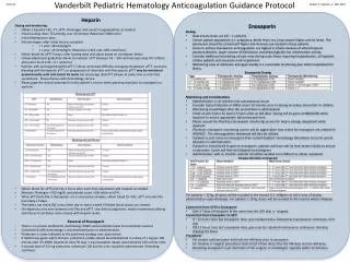
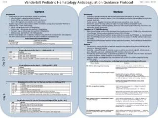
Download
1 / 34
381 likes | 1.2k Vues
Pediatric Hematology. Dr. Mariana Silva, MD.F.R.C.P.C. March 2008. FETAL AND NEONATAL ERYTHROPOIESIS. TABLE 1. Globin-chain development and composition. a This tetramer may be an epsilon tetrad.

E N D
Pediatric Hematology Dr. Mariana Silva, MD.F.R.C.P.C. March 2008
FETAL AND NEONATAL ERYTHROPOIESIS TABLE 1. Globin-chain development and composition aThis tetramer may be an epsilon tetrad. bFetal hemoglobin produced by adults has a different amino acid heterogeneity of the gamma chain atthe 136 position than fetal hemoglobin
Hemoglobin Synthesis inFetus and Newborn Gower 1 and 2 - present in yolk sac - 75% of early Hgb - undetectable after week 12 Week 12 to 32 90% Hgb F
Hemoglobin Synthesis inFetus and Newborn (cont’d) “The Switch” is Hgb F to A Hgb F after week 32; by week 40 is 50-75%; by 6 month is 5-8%; by 1 year < 1% Delayed Switch (? mechanism ? Stress Erythropoiesis) . Maternal hypoxia . Small for Gestational Age . Infants of diabetic mothers
Physiological Anemia of Infancy • Gradual in Hgb after birth for 2 months, stable 2-4 month, then • Physiologic as no symptoms of hypoxia and not nutritional Fetus - 10 weeks Hgb 9 gr/dl - 22-24 week, Hgb 14 gr/dl - 32-40 week, Hgb 16 gr/dl
Physiological Anemia of Infancy (cont’d) age: blood supply to placenta Epo to maintain oxygen supply to infant Hemoglobin (Hgb) Oxygen delivery to fetus determines Hgb level at birth. Examples: • Small for gestational • Infant of diabetic mother: metabolic demands on fetus from glucose oxygen needs Hemoglobin • Infants of smokers: fetal CO oxygen available Hemoglobin to compensate • Infants gestated at altitudes: inspired oxygen by mothers Hemoglobin in newborn
Physiological Anemia of Infancy (cont’d) • Birth: Hemoglobin (Hgb) due to placental transfusion • RBC production after birth due to availability of extrauterine oxygen • 2 month of age Hgb due to RBC production, shorter life fetal RBC • Nadir at 7-9 wk of age: Hgb 11 gr./dl
Physiological Anemia of Infancy (cont’d) placental transfusion in: • placenta previa or abruptio • multiple gestation • cord clamping < 30 seconds • C-section • Cord around neck 24-32 week retics are 15% of RBC’s, at birth 7% Day 7, retics to 1%
Postnatal changes in hemoglobin and ared-blood-cell indices in term infants
Anemia of Prematurity • Premature Infant: more rapid decline and lower nadir of Hgb than term 40% infants <33 weeks show symptoms of anemia • Epo rapidly, Epo levels 50% < than adults at same Hgb level • Epo slowly as Hgb falls in premature babies Epo produced in liver
Anemia of Prematurity (cont’d) Decision to transfuse controversy for last 30 years • Anemia risk of apnea and failure to thrive. • Transfusion at predetermined Hgb level not cost effective and doesn’t apnea
Sites and Timing of Neonatal Blood Loss A Fetus 1. Internal hemorrhage 2. Fetomaternal Hemorrhage 3. Fetoplacental hemorrhage: abruption,previa, marginal sinus ,or hematoma 4. Twin-twin transfusion: chronic
Sites and Timing of Neonatal Blood Loss (cont.) BNewborn 1. Twin-Twin transfusion: acute 2. Umbilical-cord rupture or hematoma-normal or abnormal cord 3. Internal hemorrhage: Intracranial, hepatic or splenic rupture or hematoma, adrenal or retroperitoneal hematoma. Pulmonary bleed 4. Placental trapping: caesarean section, early cord clamping, precipitous delivery
Fe Deficiency Anemia • Most common etiology of anemia Lack of dietary iron • Contributing factors in children Rapid growth Insufficient Fe • Absorption • Blood loss Breast milk or formula vs. cow’s milk • Diet Cereals Meat
Fe Deficiency Anemia (cont’d) • Lab : Hemoglobin MCV Serum Fe/TIBC Serum Ferritin • Platelets frequently • “Trial of Fe” • Treatment: Oral ferrous sulfate 5-6 mg/kg of elemental Fe x day x 3 months • Side effects of Fe
Thrombopoiesis I Yolk sac phase: • small megakaryocytes by 5 week, • platelets large and hypogranular II Hepatic phase: • early stage by 6 week • Megakaryoblasts and promegakaryocytes seen • late stage: 9-11 weeks Megakaryocytes comparable to adult, but smaller
Thrombopoiesis (cont’d) III Bone Marrow phase: after 11 weeks • from 11 to 22 stable number megakaryocytes, then 22 to 40 week. Size still small, adult size by one year of age. • Newborns easily develop thrombocytopenia due to sepsis. Little is known of newborn megakaryocytopoietic-thrombopoietic capacity
WELL Large platelets Normal hemoglobin and WBC SmallPlatelets Congenital anomalies Mean corpuscular volume Consumption Synthesis Immune Congenital ITP 2° to SLE, HIV Drug Induced TAR Wiskott-Aldrich Syndrome X-linked Amegakaryocytic Fanconi anemia Maternal ITP NATP Non-immune 2B or platelet-type vWd Hereditary macrothrombocytopenia Acquired: Medications,Toxins,Radiation
ILL Fibrinogen Fibrin degradation products Large platelets Small platelets HSM Mass Synthesis Consumption Microangiopathy Hemolytic-uremic syndrome TTP Malignancy Storage disease Sequestration Disseminated intravascular coagulation Necrotizing enterocolitis Respiratory distress ThrombosisUAC Sepsis Viral infection Hemangioma Hypersplenism
Thrombocytopenia • Infant Factors Platelet < 100,000/mm3 in 80% sick infants 60% known etiology • 22% infants in NICU have platelets Approximately 25% have significant bleeding • platelets associated with: sepsis, respiratory distress, bilirubin, ventilation, asphyxia, meconium, hypothermia, pulmonary hypertension, polycythemia • Sepsis 52-77% have platelets, may be 1st manifestation. Mean duration 5 days.
CLINICAL CASES ITyler is an 11- month- old boy brought to your office by his mother who thinks he is pale and a bit irritable. He has not had any recent illnesses and is on no medications. • History of presenting complaint • Past medical history • Diet • Family History • Physical Exam
LAB Hb 82 g/L MVC 65 WBC 6.9 x 10⁹/L Platelets 540,000 x 109/L • Possible diagnosis • Treatment
CLINICAL CASES (cont’d) II Amanda is a 2-month-old girl you are seeing today for her lst immunization. Her mother reports that Amanda sleeps a lot and looks pale. She is growing well and breastfeeds vigorously. She is on no medications. • History of presenting complaint • Past medical history • diet • Family history • Physical exam
LAB 0 Hb 102 g/L MCV 84 WBC 8.3 x 10⁹/L Platelets 240,000 x 109/L • Possible diagnosis • How would you manage this patient?
Pancytopenia Reduction below normal values of 3 blood lines: *Leukocytes * Platelets * Erythrocytes Hypocellular Marrow Seen in: Constitiutional (Inherited) marrow failure syndromes Acquired Aplastic Anemia Hypoplastic Variant of Myelodysplastic Syndromes
Pancytopenia (con’t) Cellular marrow seen in: • Primary bone marrow disease as Acute Leukemia, MDS, Myelofibrosis • Secondary to systemic disease: autoimmune disease (SLE), Vitamin B12 or folate deficiency, storage disease, metastatic solid tumors, etc.
Constitutional (Inherited) Pancytopenia Syndromes Fanconi Anemia Shwachman-Diamond Syndrome Dyskeratosis Congenita Amegakaryocytic Thrombocytopenia Other Genetic Syndromes Down Syndrome Dubowitz Syndrome Seckel Syndrome Reticular Dysgenesis Schimke Immuno-Osseous Dysplasia Familial Aplastic Anemia (non-Fanconi) Pearson Syndrome Reticular Dysgenesis Noonan Syndrome
Fanconi (Aplastic) Anemia • Autosomal recessive syndrome • Hematological findings • Typical physical anomalies • Abnormal chromosomal fragility
Anomaly Skin Pigment Changes Short Stature Upper Limb Abnormalities (thumbs,hands,radi,ulnas) Hypogonadal and Genital Changes (mostly males) Other Skeletal Findings (head/face,neck,spine) Eye/lid/epicanthal fold anomalies Renal Malformations Ear Anomalies (external & internal), deafness Hip, leg, foot, toe abnormalities Gastrointestional/cardiopulmonary malformations Approximate Frequency (% of Patients) 65 60 50 40 30 25 25 10 10 10 Characteristic Physical Anomalies in Fanconi Anemia
Marrow Failure Before Age 10 Years • Thrombocytopenia often first • Granulocytopenia • Macrocytic Anemia • Severe Aplastic Anemia in Most Cases • Increased Propensity for Cancer: Carcinomas of Head, Neck, Carcinoma of Vulva and Anus
Treatment • Transfusions • Hematolpoietic Stem Cell Transplant is only Curative Treatment • Androgens – Prednisone Prognosis: Median Survival is >30 years of Age