Download
1 / 97

E N D
Nephrotic syndrome and nephritic syndrome Submitted to Dr. Digambar nartam(HOD, MD) Dr. Pawan garg(MD) Dr. Arvind tripathi(MD) SUBMITTED BY RATNESH KUMAR SHUKLA B. A. M. S. Final year
Reference :- • KUMAR AND CLARKS CLINICAL MEDICINE, 9TH EDITION, CHAPTER 20-KIDNEY AND URINARY TRACT DISEASE • HARRISONS PRINCIPLE OF INTERNAL MEDICINE 20TH EDITION • API TEXTBOOK OF MEDICINE, SECTION 19 • TEXTBOOK OF PATHOLOGY, BY HARSHMOHAN • ROBINS BASIC PATHOLOGY • ESSENTIAL OF MEDICAL PHYSIOLOGY 6TH EDITION, BY K SEMBULINGAM • www.wikipaedia.com • www.ncbi.nlm.nih.gov
NEPHRON • A nephron is the basic unit of structure and function in the kidney. A nephron is used separate to water, ions and small molecules from the blood, filter out wastes and toxins, and return needed molecules to the blood. • Each nephron has two major portions: • A Renal corpuscle (Malpighian body) • A Renal tubule
Glomerulus • The glomerulus is a tuft of small blood vessels called capillaries located within Bowman's capsule within the kidney. • Glomerular mesangial cells structurally support the tufts. • Blood enters the capillaries of the glomerulus by a single arteriole called an afferent arteriole and leaves by an efferent arteriole.
Structure of Glomerular wall Glomerular Capillary wall is filtering area. • It consists of three basic structure • The layer of fenestrated endothelium • Glomerular basement Membrane • Visceral epithelial layer(podocytes)
GLOMERULAR FILTRATION BARRIER • Urine formation begins at the glomerular filtration barrier. • Theglomerular filter through which the ultrafiltrate has to pass consists of three layers: the fenestrated endothelium, the interveningglomerular basement membrane, and the podocyte slit diaphragm. • There are two kindkinds of barrier for filtration . • Size barrier • Ion barrier
2. Ion barrier • There is another barrier called as negative charges barrier. • GBM, endothelium and podocyte are negatively charged. • These negetive Charged layers address to the repulsion for negetively charged molecule. • Ex. – plasma protein at normal physiological pH charged negetively like albumin.
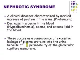
NEPHROTIC SYNDROME • Nephrotic syndrome refers to clinical condition that includes five main characters :- • Massive proteinuria (more then 3.5gm/day) • Hypo albuminaemia (less then 3gm/dl) • Generalized edema • Hyperlipidaemia (increase cholesterol and triglycerides) • lipidurea
1.proteinurea • Proteinuria occurs partly because structural damage to the glomerular barrier (podocytes, membrane, fenestrated endothelium and endothelial charge) allows the passage of more and molecules. • Protein leaked out more then 3.5gm/day.(nephrotic range proteinurea) • The filtration slit between podocytes and normal podocyte architecture, and Podocyte foot processes are critical to maintaining a barrier to protein loss into the urinary space, as is Functional GBM and healthy capillary endothelium (and its charge).I • Proteinurea maybe in two types :- • Selective proteinuria (only albumin leaked out) • Non-selective proteinuria
2.Hypoalbuminaemia • Loss of urinary protein (largely albumin) of the order ≥3.5 g daily in an adult may lead to hypoalbuminaemia. • the normal liver can synthesize albumin at a rate of 10–12 g daily. • This can be partly explained by increased catabolism of reabsorbed protein, largely albumin, in the proximal tubules, even though the rate of albumin synthesis is increased. • Loss of plasma protein in urine can decrease level of oncotic pressure in circulatory compartment and results as anasarca.
3.Hyperlipidaemia • This is a consequence of increased synthesis of lipoproteins as a direct consequence of a low plasma albumin. • Low-density lipoprotein (LDL) increases, Very-low-density lipoprotein (VLDL) and/or intermediate-density lipoprotein (IDL) fractions increase, with no change (or a decrease) in HDL. Hyperlipidaemia is caused by two factors: • Hypoproteinemia stimulates protein synthesis in the liver, resulting in the overproduction of lipoproteins. • Lipid catabolism is decreased due to lower levels of lipoprotein lipase, the main enzyme involved in lipoprotein breakdown.
4. Edema • Edema is thought to be caused by two mechanisms. • The first being hypoalbuminemia which lowers the oncotic pressure within vessels resulting in hypovolemia and subsequent activation of the renin–angiotensin system and thus retention of sodium and water. • Nephrotic syndrome edema initially appears in parts of the lower body (such as the legs) and in the eyelids. In the advanced stages it also extends to the pleural cavity and peritoneum (ascites) and can even develop into a generalized anasarca. • Edema maybe appear more in most dependent area like, walking-legs, sleeping-back.
5.Lipidurea • Lipiduria or loss of lipids in the urine is indicative of glomerular pathology due to an increase in the filtration of lipoproteins • When Lipid passed through nephrons tubules some cells of proximal convulated tubes started pinocytosis and become globulated. • These cells become dysfunctional and shut down in urine. • In the urine analysis process may be found big fat globules they called as “fat ovale bodies”.
Causes of Nephrotic syndrome • Nephrotic syndrome may be:- • Primary • Secondary 2.Secondery causes :- • DM, • Amyloidosis • Drugs (gold, penicillamine) • Infection (malaria, syphilis,HIV,… ..) • Primary causes – • Minimal change disease • Membranous glomerulopathy • Focal segmental glomerulosclerosis • Membranousproliferative glomerulopathy
1.Minimal changes disease • the most common cause of nephrotic syndrome in children. • It owes its name to the fact that the nephrons appear normal when viewed with an optical microscope as the lesions are only visible using an electron microscope. • Another symptom is a pronounced proteinuria. • The individual foot processes can no longer be made out- it is like they have all just “melted” together into a single thin layer. This important barrier in the filtration process can no longer keep protein from being filtered out of the blood and into the urine.
Many drugs have been implicated in MCN, including NSAIDs, lithium, antibiotics (cephalosporins, rifampicin, ampicillin), bisphosphonates and sulfasalazine. • Atopy is present in 30% of cases of MCN, and allergic reactions can trigger the nephrotic syndrome. • Infections, such as hepatitis C virus (HCV), the human immunodeficiency virus (HIV) and tuberculosis, are rarer causes.
Clinical feature • MCN is most common in children, particularly boys, and is responsible for the large majority of cases of nephrotic syndrome in childhood. • Proteinuria is usually ‘highly selective’, where albumin, but not highermolecular-weight proteins such as immunoglobulins, is lost in the urine. • Oedema is usual and in children this may present predominantly around the face.
Management Manage symptoms with general measures (above). •The first line therapy to minimal change disease is corticosteroids. High-dose corticosteroid therapy with prednisolone 80mg/day daily • In children, two-thirds relapse after steroid therapy and further courses of corticosteroids are required. One-third of these children regularly relapse on steroid withdrawal, so that a second-line agent shouldbe added after repeat induction with steroids. • Cyclophosphamide 1.5–2.0 mg/kg daily is given for 8–12 weeks with prednisolone 7.5–15 mg/day. This increases the likelihood of long-term remission. Steroid-unresponsive patients may also respond tocyclophosphamide. No more than two courses of cyclophosphamide should be prescribed in children because of the risk of side-effects, which include future infertility (azoospermia and premature ovarian failure).
2.Membranous glomerulopathy • Idiopathic membranous glomerulopathy is an autoimmune disease that occurs mainly in adults, and predominantly in men. • Microscopic haematuria, hypertension and renal impairment may accompany the nephrotic syndrome. • As in other glomerular disease, hypertension and a greater degree of renal impairment are poor prognostic signs. • Membranous nephropathy is considered an autoimmune disease, which means that it caused by the body’s own immune system. MN is caused by the build-up of immune complexes within the filters (glomeruli) of the kidney itself. • antibodies and antigens create immune complexes that get stuck in the kidney filter (glomerulus) and cause disease. • These autoantibodies are Anti pla2r (anti-phospholipase A2 receptor antibody) and Anti thsd7a(anti-thrombospondin type 1 domain-containing 7A) • Doctors can be diagnosed differ kind of nephrotic syndrome by biopsy of kidney.
Causes of membranous glomerulopathy In the primary or idiopathic form (which comprises 75% of the cases), glomerular histology is identical to that seen when membranous glomerulopathy is secondary to another insult. These include: • drugs (e.g. penicillamine, gold, NSAIDs, probenecid, mercury, captopril) • autoimmune disease • infections • cancers • other causes
Management • patients should receive ACE inhibition . • The alkylating agents, cyclophosphamide (1.5–2.5 mg/kg per day for 6–12 months with 1 mg/kg per of oral prednisolone on alternate days for the first 2 months) and chlorambucil (0.2 mg/kg per day in months 2, 4 and 6, alternating with oral prednisolone 0.4 mg/kg per day in months 1, 3 and 5), are effective. • Mycophenolate mofetil has demonstrated benefit in smaller studies with short follow-up. • Anti-CD20 antibodies (rituximab, which ablates B lymphocytes) have been shown to improve renal function, reduce proteinuria and increase the serum albumin; no significant adverse affects have been shown in the short term. • Oral corticosteroids are of no benefit alone but may be additive. A pilot study has shown promise with subcutaneous administration of adrenocorticotrophic hormone (tetracosactide) twice weekly, demonstrating an improvement in proteinuria. It is believed that it acts directly on podocytes by binding to melanocortin receptors. It is licensed by the FDA for use in nephrotic syndrome of any cause.
3.Focal segmental glomerulosclerosis • Focal segmental glomerulosclerosis (FSGS) describes a sclerotic glomerular lesion that affects some (but not all) glomeruli, and some (but not all) segments of each tuft. • hypoalbuminaemia is unusual. • Immunofluorescence description • IgM and C3 deposited in focal and segmental manner in the sclerotic segments • Electron microscopy description • Epithelial cell detachment from glomerular basement membrane • Extensive foot process obliteration (even in nonsclerotic glomeruli), mesangial sclerosis with increased matrix and collapsed glomerular loops
A. Primary FSGS • This disease of unknown aetiology usually presents as massive proteinuria (usually non-selective),haematuria, hypertension and renal impairment. • The associated nephrotic syndrome is often resistant to steroid therapy. All age groups are affected. • It usually recurs in transplanted kidneys, sometimes days of transplantation, and particularly in patients with aggressive primary renal disease.
B. Secondary fsgs • Secondary(Secondary, when an underlying cause is identified) FSGS with similar glomerular changes is seen as a secondary phenomenon when the Number functioning nephrons is reduced for any reason. • As nephrons fail, increased flow through the remaining nephrons leads to glomerular hypertrophy and hyperfiltration With the secondary changes of FSGS. This is actually including numerous causes such as • Toxins and drugs such as heroin and pamidronate. • diabetes mellitus • Kidney defects from birth • Urine backing up into kidneys • Obesity • Sickle Cell Anemia • Viruses (such as HIV)
Management • Medications that suppress your immune system • Diuretics and low salt diet help to control edema • A medication that blocks a hormone system called the renin angiotensin system (ACE inhibitor or ARB) to control blood pressure or lower urine protein • Anticoagulants to prevent blood clots • Statins to lower the cholesterol level Prednisolone 0.5–2 mg/kg per day is used in most patients and continued for 6 months • Ciclosporin at doses to stopping urinary protein excretion (tacrolimus is an alternative). Relapse after reducing or stopping ciclosporin is very common so that long-term use is required. • Cyclophosphamide, chlorambucil or azathioprine is used for second-line therapy in adults.
4.Membranoproliferative glomerulopathy • Membranoproliferative glomerulonephritis (MPGN) is an uncommon cause of chronic nephritis that occurs primarily in children and young adults. This entity refers to a pattern of glomerular injury based on the following three characteristic histopathologic findings: • Proliferation of mesangial and endothelial cells and expansion of the mesangial matrix • Thickening of the peripheral capillary walls by subendothelial immune deposits and/or intramembranous dense deposits • Mesangial interposition into the capillary wall, giving rise to a double-contour on light microscopy
Three variations are founded Type III – This is also an immune complex disease, similar to Type I. However, the immune complexes are found in the subepithelial space, and there is disruption of the glomerular basement membrane with large open areas. Type II – This is also called dense deposit disease. When viewed under the microscope, continuous, dense ribbon-like deposits are found along the basement membranes of the glomeruli, tubules, and Bowman’s capsule. • Type I – Discrete immune complexes are found in the mesangium and subendothelial space. Immune complexes are combinations of antigens and antibodies which bind to each other and then become lodged in the kidney. This activates the immune system, which causes inflammation and damage to the kidney itself.
Management • In idiopathic MCGN (all age groups) with normal renal function and non-nephrotic-range proteinuria, No specific therapy is required. Good blood pressure control, ideally with an ACE inhibitor, is of benefit. • If no benefit is seen, this treatment is discontinued. Regular follow-up, with control of blood pressure, use of agents to reduce proteinuria and correction of lipid abnormalities, is necessary.
Congenital Nephrotic syndrome • Congenital nephrotic syndrome (Finnish type) is an autosomal recessively inherited disorder due to mutations in the gene coding for a transmembrane protein, nephrin; . Nephrin is a critical element of the filtration slit, and its loss of function results inmassive proteinuria shortly after birth. • The disorder can be diagnosed in utero, as increased α-fetoprotein in amniotic fluid is a common feature. • Histologically, some glomeruli are small and infantile, whereasothers are enlarged and more mature, and have diffuse mesangial hypercellularity. Because of the massive • proteinuria, some tubules develop microcysts and are dilated. On electron microscopy, complete effacement of the foot processes of visceral epithelial cells is observed. This condition is characterized by relentless progression to ESKD.
Treatment • Early and aggressive treatment is needed to control this disorder. • Treatment may involve: • Antibiotics to control infections • Blood pressure medicines called angiotensin-converting enzyme (ACE) inhibitors and angiotensin receptor blockers (ARBs) to reduce the amount of protein leaking into the urine • Diuretics ("water pills") to remove excess fluid • NSAIDs, such as indomethacin, to reduce the amount of protein leaking into the urine • Fluids may be limited to help control swelling. • The provider may recommend removing the kidneys to stop protein loss. This may be followed by dialysis or a kidney transplant.
Secondery causes of NS :- HIV associated nephrotic syndrome • In HIV-associated nephropathy (HIVAN), glomeruli are characteristically ‘collapsed’ on light microscopy podocytes are enlarged, hyperplastic and coarsely vacuolated, containing protein absorption droplets and overlying capillaries with varying degrees of wrinkling and collapse of the walls. • Direct podocyte HIV-1 infection is associated with loss of podocyte-specific markers such Wilms’ tumour factor and synaptopodin in HIVAN. • HIVAN presents with nephrotic-range proteinuria, edema and CKD, which can be rapid in progression. Antiretroviral therapy (ART) may reverse the reanal lesions seen, and restores renal function if treatment is commenced early
Management • Treatment with highly active antiretroviral therapy and angiotensin converting enzyme inhibitors or angiotensin receptor blockershas been shown to be beneficial and should be given to all patients unless otherwise contraindicated. General renoprotective measures and the treatment of the complications of nephrotic syndrome and kidney failure are adjunctive. • Corticosteroid treatment can be useful in patients who do not respond to initial treatment. There is some evidence that ciclosporin might be helpful in selective cases, however further study of both steroids and ciclosporin is needed before these types of drugs can be considered standard treatment.
Amyloidosis • Amyloidosis is a systemic acquired or inherited disorder of protein folding, in which normally soluble proteins or fragments are deposited extracellularly as abnormal insoluble fibrils,causing progressive organ dysfunction and death. • The abnormal protein may be derived from light chains or • immunoglobulin (AL amyloid), or Serum amyloid A protein (AA amyloid). The renal consequences are similar, even if systemic features Differ.
Management • Treatments that reduce production of the amyloidogenic protein can improve organ function and survival in immunoglobulin-light-chain-related (AL) amyloidosis and hereditary transthyretin-associated (ATTR) Amyloidosis . • In AA amyloidosis, production of serum amyloid A can sometimes be decreased by treatment of the underlying inflammatory condition but cannot be completely suppressed. • Renoprotective measures should be started. The success of dialysis and kidney transplantation depends on the extent of amyloid deposition in extrarenal sites, especially the heart.
Diabetic nephropathy • Diabetic renal disease is the leading cause of ESKD in the Western world, arising largely as acomplication of type 2 diabetes mellitus. • Diabetic kidney disease occurs in about 20–30% of both type 1 and type 2 diabetics the natural history is similar from the onset of proteinuria, and the histological lesion is the same. Pathology • there is constriction of the efferent arterioles and dilation of afferent arterioles, with resulting glomerular capillary hypertension and hyperfiltration. • Glomerular hyperfiltration (the GFR increases to >150 mL/min/m2 ) and initial enlargement of kidney volume occur as local vasoactive factors increase flow. The GBM thickens and the mesangium expands.Progressive depletion of podocytes from the filtration barrier (through apoptosis or detachment) results in podocyturia early in the disease. Proteinuria evolves as filtration pressures rise and the filter is compromised. Later, glomerulosclerosis develops with nodules (Kimmelstiel–Wilson lesion - matrix invades the glomerular capillaries and produces deposits called Kimmelstiel-Wilson nodules) and hyaline deposits in the glomerular arterioles .
Management Lifestyle changes (cessation of smoking, attention to salt intake, weight loss and increased exercise) are necessary in preventing progression of any diabetic complication. • Aim for good (intensive) glycaemic control. If achieved for even a limited period, this reduces the incidence of ESKD and other microvascular complications in the long term (the so-called ‘legacy effect’. • Control dyslipidaemia. • Control blood pressure to <120/80 mmHg with ACE inhibitors or AII-RA; these should be used once microalbuminuria develops, even if blood pressure control is good. • Combined use of ACE inhibitors and AII-RA (dual blockade) does not provide additional benefit but is associated with an increased risk of AKI and hyperkalaemia; it is no longer recommended in recent trials.
Other related nephropathy • Systemic lupus erythematosus: This autoimmune disease can affect a number of organs, among them the kidney, due to the deposit of immunocomplexes that are typical to this disease. The disease can also cause lupus nephritis. • Sarcoidosis: This disease does not usually affect the kidney but, on occasions, the accumulation of inflammatory granulomas(collection of immune cells) in the glomeruli can lead to nephrotic syndrome. • Syphilis: kidney damage can occur during the secondary stage of this disease (between 2 and 8 weeks from onset). • Hepatitis B: certain antigens present during hepatitis can accumulate in the kidneys and damage them. • Sjögren's syndrome: this autoimmune disease causes the deposit of immunocomplexes in the glomeruli, causing them to become inflamed, this is the same mechanism as occurs in systemic lupus erythematosus.