Download
1 / 5
50 likes | 60 Vues
To investigate the inhibition of cisplatin-resistant tumor cells on activated CD4 T cells in ovarian cancer and its mechanism.

E N D
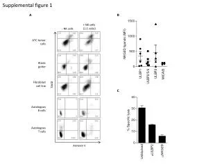
Pharmaceutical Research Article Inhibition of cisplatin-resistant tumor cells on activated CD4+ T cells in ovarian cancer and its mechanism Objective: To investigate the inhibition of cisplatin-resistant tumor cells on activated CD4+ T cells in ovarian cancer and its mechanism. Li H*, Tian S, Liu Z, Yu J & Yan J Methods: 50 patients with ovarian cancer admitted to our hospital from March 2016 to July 2017 were selected as the research group, and 50 normal subjects were selected as the control group. The same separation method of CD4+ T cells was applied to both two groups. The serum levels of ovarian cancer markers and inflammation factors were compared between two groups. Qilu Hospital of Shandong University, Infertility Center, Jinan, China*Author for Correspondence: tu41267@163.com Results: The serum levels of ovarian cancer markers in the research group were higher than those in the control group (P<0.05). The average proportion of CD4+ T cells in peasant patients was higher than that in workers and cadres (P<0.05). The average proportion of CD4+ T cells in patients with tumor infiltration was higher than that in patients without infiltration (P<0.05). The level of IL-6 in the research group was higher than that in the control group. While the levels of IL-10, IL-4, IL-2, TNF-α and IFN-γ were lower than those in the control group (P<0.05), and the immune function of the research group was still lower than that of the control group (P<0.05). Conclusion: Cisplatin-resistant ovarian cancer cells can inhibit the activation of CD4+ T cells, which can effectively judge the different stages of ovarian cancer. Moreover, the number of CD4+ T cells can be used to determine the prognosis of patients, providing a certain clue for clinical treatment. Keywords:ovarian cancer ▪ cisplatin resistant tumor cells ▪ activated CD4+ T cells ▪ inhibition Healthy resist the invasion of germs, clear its own mutant cells, and maintain the balance of autoimmune function [3]. However, tumors are caused by the fact that the mutant cells are not effectively cleared and continue to propagate. CD4+ T cells belong to the inhibitory immunoregulatory cells, which play a very important role in the human immune response and also play an inhibitory role in the process of resisting tumors, so as to avoid the escape of tumor cells [4]. The inhibition of cisplatin-resistant tumor cells on activated CD4+ T cells and its mechanism were investigated in our hospital. It is believed that cisplatin-resistant tumor cells have inhibitory effects on activated CD4+ T cells, and the report is as follows. Materials and methods human body can effectively Introduction Ovarian cancer is one of the female malignant tumors, which is mainly found in the female reproductive system. The initial symptoms of most patients are not obvious, and they are in advanced stages by the time they are diagnosed, which has a negative impact on women’s life and health [1]. The main method for clinical treatment of ovarian cancer is chemotherapy. The chemotherapeutic drugs are mostly platinum-based drugs Cis- Diaminedichloroplatinum (CDDP), but the toxic side effects of chemotherapy on patients are very large, and long-term treatment of tumors will also produce drug resistance, which seriously reduces the therapeutic effect [2]. When tumor cells become resistant to drugs, many factors will be produced to change the tumor microenvironment, and the immune response will be inhibited to accelerate the development of the disease. General data 154 Pharm. Bioprocess. (2018) 6(4), 142–153 ISSN 2048-9145
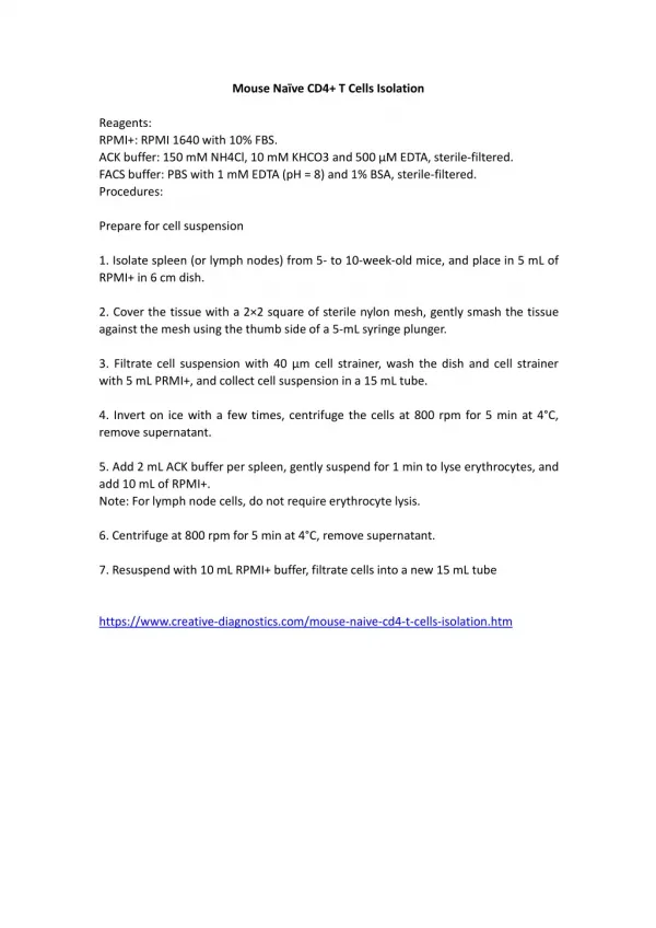
Research Article Li et al., 10 min, and the separated serum was placed in a refrigerator at -80°C. ELISA was used to detect IL-4, IL-10, IL-6, IL-2 and so on. The kit was purchased from Shanghai Kanglang Biotechnology Co., Ltd. and was performed according to the operating instructions. The CRP was measured using an automatic biochemical analyzer. 50 patients with ovarian cancer admitted to our hospital from March 2016 to July 2017 were selected as the research group, and 50 normal subjects were selected as the control group. Inclusion criteria: 1) 50 cases of patients with ovarian cancer were diagnosed by cytology and pathology; 2) the two groups of personnel were informed and agreed with this study. Statistical methods SPSS 18.0 statistical software was used for data analysis and processing. The count data were determined by chi-square test (%), while the measurement data were expressed as mean ± standard deviation, and the t test was used. P<0.05 means a significant difference. Results • Comparison of serum ovarian cancer markers between the two groups Exclusion criteria: 1) patients with other tumors; 2) patients with infectious diseases. The two groups were female, and the control group was 25-76 years old with an average age of (66.2 ± 4.1) years. The research group was 23-75 years old, with an average age of (67.3 ± 3.5) years. The general data of the two groups were comparable, and the study was approved by the hospital ethics committee. The serum ovarian cancer markers in the research group were higher than those in the control group (P<0.05), as shown in TABLE 1. Methods The same CD4+ T cell separation method was applied to both two groups, namely MACS magnetic separation [5]. The peripheral blood of the two groups was extracted for 20 ml, and the aseptic operation was ensured during the extraction process. The mononuclear cells were subjected to density gradient centrifugation, and the CD4+ T cells were separated by MACS magnetic separation column. A small amount of the cell suspension was subjected to purity detection by flow cytometry, and activation was carried out only when the purity was >90%, and the activated antibody was anti-CD28 and anti-CD3 antibodies, followed by continuous activation for 3 days [6]. • Analysis CD4+ T cells and clinical pathological parameters in patients with ovarian cancer of relationship between • The average proportion of CD4+ T cells in patients with ovarian cancer older than 40 years old was higher than that in patients with ovarian cancer less than 40 years old (P<0.05). The average proportion of CD4+ T cells in peasant patients was higher than that in workers and cadres (P<0.05). Moreover, the average proportion of CD4+ T cells in serous adenocarcinoma was higher than that of mucinous adenocarcinoma and mixed type (P<0.05). The average proportion of CD4+ T cells in patients with stage IV ovarian cancer was higher than that in patients with stage I-III (P<0.05), and the average proportion of CD4+ T cells in ovarian cancer patients without metastasis was higher than that in patients with metastasis (P<0.05). Besides, the average proportion of CD4+ T cells in patients with tumor infiltration was higher than that in patients without infiltration (P<0.05). The average proportion of CD4+ T cells in patients with poorly differentiated ovarian cancer was higher than that in moderately differentiated and highly Observational index (1) The serum levels of ovarian cancer markers were compared between the two groups, including cancer antigen 125 (CA125), Human epididymis protein 4 (HE4), and vascular endothelial growth factor (VEGF). (2) The relationship between CD4+ T cells and clinical pathological parameters in patients with ovarian cancer was analyzed. (3) The levels of IL-10, IL-4, IL-6, IL-2, TNF-α, IFN-γ and other cytokines in plasma were compared between the two groups. (4) The immune function of the two groups was compared. Elisa assay Total 3 ml venous blood after 2 weeks treatment with cisplatin was taken in the morning, centrifuged at 2000 rpm/min for Pharm. Bioprocess. (2018) 6(4) 155
Research Article Inhibition of cisplatin-resistant tumor cells on activated CD4+ T cells in ovarian cancer and its mechanism Table 1. Comparison of serum ovarian cancer markers between the two groups Groups Cases CA125 (U/ml) Control group 50 20.4 ± 3.3 Research group 50 120.4 ± 24.2 T / 12.383 P / <0.05 HE4 (pmol/L) 73.5 ± 8.4 176.3 ± 23.5 16.184 <0.05 VEGF (pg/ml) 110.3 ± 13.2 239.3 ± 26.2 21.677 <0.05 inhibit their immune function [12]. The number of CD4+ T cells in peripheral blood of patients with ovarian cancer is relatively high. The increase of the number of CD4+ T cells in patients with ovarian cancer will inhibit the immune response, weaken the ability of immune cells to resist tumor and promote the growth and reproduction of tumor cells [13]. It was demonstrated in the results that the average proportion of CD4+ T cells in patients with ovarian cancer older than 40-year-old was higher than that in patients with ovarian cancer less than 40 years old (P<0.05). The average proportion of CD4+ T cells in peasant patients was higher than that in workers and cadres (P<0.05). Moreover, the average proportion of CD4+ T cells in serous adenocarcinoma was higher than that of mucinous adenocarcinoma and mixed type (P<0.05). The average proportion of CD4+ T cells in patients with stage IV ovarian cancer was higher than that in patients with stage I-III (P<0.05), and the average proportion of CD4+ T cells in ovarian cancer patients without metastasis was higher than that in patients with metastasis (P<0.05). Besides, the average proportion of CD4+ T cells in patients with tumor infiltration was higher than that in patients without infiltration (P<0.05). proportion of CD4+ T cells in patients with poorly differentiated ovarian cancer was higher than that in moderately differentiated and highly differentiated patients (P<0.05). The above results suggested that the more CD4+ T cells, the more CD4+CD25+Treg content, which is conducive to promoting the development and occurrence of tumors. Meanwhile, the immune ability of the body will significantly decline after the occurrence of the activation of CD4+ T cells, thereby increasing the incidence of tumors. During the development of ovarian cancer, cancer cells can secrete a variety of specific factors. By detecting the contents of ovarian cancer marker in the patient’s serum, the progression of the tumor can differentiated patients (P<0.05). The specific details were shown in TABLE 2. • Comparison of plasma levels of IL-10, IL-4, IL-6, IL-2, TNF-α, IFN-γ and other cytokines between the two groups • The level of IL-6 in the research group was higher than that in the control group. While the levels of IL-10, IL-4, IL-2, TNF-α and IFN-γ were lower than those in the control group (P<0.05), as demonstrated in TABLE 3. • Comparison between the two groups of immune function The immune function of the research group was lower than that of the control group (P<0.05), as shown in TABLE 4. Discussion Ovarian cancer has a high mortality rate, ranking among the top three in female malignant tumors, and it is already in advanced stage because it has no abnormal symptoms in the early stage [7,8]. The main method of clinical treatment is chemotherapy, but the emergence of drug resistance after chemotherapy is the cause of treatment failure. At the same time, the metastatic ability of drug-resistant cells is very strong, which can induce other cytokines to produce immunosuppressive effect and reduce the ability of T cells to clear abnormal cells [9]. The average The immune escape of the body is the main cause of ovarian cancer. Under normal circumstances, the immune cells of the human body will recognize and clear the abnormal proliferating cells, thereby effectively preventing the normal cells from developing lesions and reducing the incidence of tumors [10]. If the immune function of the human body is inhibited or is not working properly, the immune cells cannot recognize and clear the abnormal cells, thereby promoting the development of the tumor [11]. CD4+ T cells are a subset of immunosuppressive cells. CD4+CD25+Treg cells generated by activated CD4+ T cells can inhibit the activation process of T cells and 156
Research Article Li et al., Table 2. Analysis of relationship between CD4+ T cells and clinical pathological parameters in patients with ovarian cancer (Cases, %) Items Cases Age >40 43 20.2 ± 6.8 <40 7 18.1 ± 8.2 Occupation Peasant 15 23.3 ± 7.2 Worker 12 22.6 ± 5.2 Cadres 23 17.8 ± 3.6 Histopathology Serous adenocarcinoma 37 22.6 ± 7.1 Mucinous adenocarcinoma Mixed type 5 17.2 ± 5.3 Staging I 10 16.8 ± 4.4 II-III 26 19.1 ± 6.1 IV 14 27.6 ± 4.1 Metastasis With 35 20.4 ± 6.4 Without 15 22.2 ± 7.3 Infiltration With 39 22.5 ± 6.4 Without 11 17.8 ± 3.2 Differentiated degree Highly differentiated 8 15.1 ± 0.9 Moderately differentiated 12 20.7 ± 6.4 Poorly differentiated 30 24.1 ± 5.8 x ± s 95% CI 19.1-24.1 6.2-32.1 20.3-28.1 17.1-28.8 13.6-23.4 20.8-26.1 P <0.05 <0.05 <0.05 <0.05 <0.05 <0.05 <0.05 8 21.0 ± 6.0 14.5-28.5 <0.05 14.4-21.2 13.6-21.5 16.6-22.4 25.1-31.2 18.8-24.3 18.1-24.2 19.8-26.4 10.5-27.1 13.1-18.1 15.4-27.3 20.9-28.3 <0.05 <0.05 <0.05 <0.05 <0.05 <0.05 <0.05 <0.05 <0.05 <0.05 Table 3. Comparison of plasma levels of IL-10, IL-4, IL-6, IL-2, TNF-α, IFN-γ and other cytokines between the two groups ( x s t Groups Cases IL-10 (pg/ml) ml) Control group Research group T / 12.367 15.746 P / <0.05 <0.05 ) ± IL-4 (pg/ IL-6 (pg/ ml) IL-2 (pg/ ml) TNF-α (pg/ ml) IFN-γ (pg/ ml) 50 4.3 ± 2.2 5.5 ± 3.4 5.0 ± 2.5 2.4 ± 1.1 3.1 ± 1.2 18.9 ± 4.4 50 3.9 ± 2.1 4.7 ± 2.8 86.1 ± 55.8 0.9 ± 0.3 1.0 ± 1.1 7.4 ± 2.1 21.466 <0.05 13.450 <0.05 16.233 <0.05 18.146 <0.05 Table 4. Comparison of immune function between the two groups ( Groups Cases Control group 50 Research group T / P / ) ± x s CD3+/CD4+ 36.3 ± 6.9 CD3+/CD8+ 27.6 ± 5.7 CD4+/CD8+ 1.2 ± 0.3 50 41.8 ± 6.3 25.1 ± 6.2 1.6 ± 0.6 19.356 <0.05 15.560 <0.05 12.259 <0.05 be effectively judged [14]. The markers for ovarian cancer are mainly CA125, HE4, and VEGF [15]. It was exhibited in the studies that the serum levels of ovarian cancer markers in the research group were higher than those in the control group (P<0.05). From this, it can be found that the serum levels of CA125, HE4, and VEGF in patients with ovarian cancer are significantly higher than those in normal subjects. Moreover, the levels of CA125, HE4 and VEGF in patients were proportional to the number of CD4+ T cells. To sum up, cisplatin-resistant ovarian cancer Pharm. Bioprocess. (2018) 6(4) 157
Research Article Li et al., cells can inhibit the activation of CD4+ T cells, which can effectively judge the different stages of ovarian cancer. Moreover, the number of CD4+ T cells can be used to determine the prognosis of patients, providing a certain clue for clinical treatment. References 7. Krishnamurthy A, Jimeno A. Bispecific antibodies for cancer therapy: a review. Pharmacology & therapeutics. Pharmacol. Ther. 185, 122-134 (2017). 8. Markowska A, Sajdak S, Huczyński A, et al. Ovarian cancer stem cells: A target for oncological therapy. Adv. Clin.Exp. Med. (2018). 9. Maschmeyer P, Zimmermann J, Tran CL, et al. A7. 19 Systemic inhibition of MIR-148A by antagomirs reduces CD4+ T helper cell numbers and alleviates inflammation in a preclinical model of transfer colitis. Ann. Rheu. Dis. 75, A63 (2016). Kim JE, Jang MJ, Jin DH, et al. Paclitaxel- exposed ovarian cancer cancer-specific CD4+ T cells after doxorubicin exposure through regulation of MyD88 expression. Int. J. Oncol. 44(5), 1716-1726 (2014). 1. cells induce 10. Zhou J, Li L, Wang L, et al. Establishment of a SVM classifier to predict recurrence of ovarian cancer. Mol. Med. Rep. (2018). Wu W, Liu Y, Ye H, et al. Millepachine showed novel antitumor effects in cisplatin‐resistant human ovarian cancer through inhibiting drug efflux function of ATP‐binding cassette transporters. Phytother. Res. (2018). 2. 11. Behbakht K, Cohn DE, Straughn Jr JM. Hyperthermic intraperitoneal chemotherapy (HIPEC) is cost-effective in the management of primary ovarian cancer. Gynecol. Oncol. (2018). Wu Z, Mei X, Ying Z, et al. Ultraviolet B inhibition of DNMT1 activity via AhR activation dependent SIRT1 suppression in CD4+ T cells from systemic lupus erythematosus patients. J. Dermatol. Sci. 86(3), 230-237 (2017). 3. 12. Ma K, Ma P, Lu H, et al. Fentanyl suppresses the survival of CD 4+ T cells isolated from human umbilical cord blood through inhibition of IKK s‐mediated NF‐κB activation. Scandinavian. J. Immunol. 85(5), 343-349 (2017). Yang L, Wang S, Zhang Q, et al. Clinical significance of immune microenvironment in ovarian cancer patients. Mol. Omics. (2018). 4. 13. Stoelting DP, Borrmann M, Koch M, et al. How liposomal cisplatin overcomes chemoresistance in ovarian tumour cells. Anticancer. Res. 34(1), 525-530 (2014). 5. Alsamman K, El-Masry OS. Staurosporine alleviates cisplatin human cancer cell models by suppressing the induction of SQSTM1/p62. Oncol. Rep. 40(4), 2157-2162 (2018). chemoresistance in 14. Yu PN, Yan MD, Lai HC, et al. Downregulation of miR‐29 contributes to cisplatin resistance of ovarian cancer cells. Int. J. Cancer. 134(3), 542-551 (2014). 6. Wojas-Turek J, Szczygieł A, Kicielińska J, et al. Treatment with cyclophosphamide supported by various dendritic cell-based vaccines induces diversification in CD4+ T cell response against MC38 colon carcinoma. Int. J. Oncol. 48(2), 493-505 (2016). 15. Jian Z, Jing L, Chen H, et al. Specific immunotherapy generates CD8 +, CD196 +, T cells to suppress lung cancer growth in mice. Immunol. Res. 64(4), 1033-1040 (2016). Pharm. Bioprocess. (2018) 6(4) 158