THE LIGHT MICROSCOPE AND THE ELECTRON MICROSCOPE
THE LIGHT MICROSCOPE AND THE ELECTRON MICROSCOPE. © 2007 Paul Billiet ODWS. UNITS OF MEASUREMENT. 1m = 10 3 mm (millimetres) 1m = 10 6 µm (micrometres) 1m = 10 9 nm (nanometres) Sometimes in old texts Angstroms (Å) are used (the diameter of a hydrogen atom) 1m = 10 10 Å .

THE LIGHT MICROSCOPE AND THE ELECTRON MICROSCOPE
E N D
Presentation Transcript
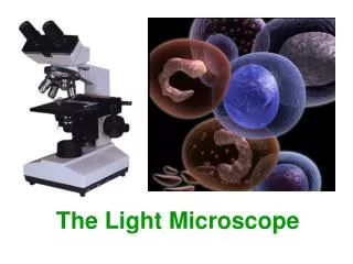
THE LIGHT MICROSCOPE AND THE ELECTRON MICROSCOPE © 2007 Paul Billiet ODWS
UNITS OF MEASUREMENT 1m = 103 mm (millimetres) 1m = 106 µm (micrometres) 1m = 109 nm (nanometres) Sometimes in old texts Angstroms (Å) are used (the diameter of a hydrogen atom) 1m = 1010 Å © 2007 Paul Billiet ODWS
The electromagnetic spectrum © 2007 Paul Billiet ODWS
Light v Electron © 2007 Paul Billiet ODWS
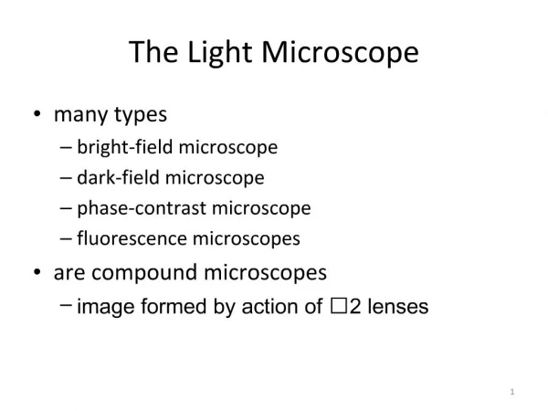
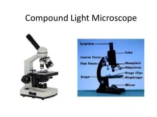
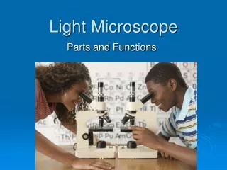
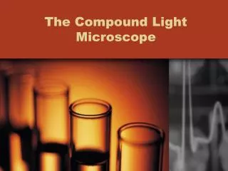
Magnifying using the Light Microscope © 2007 Paul Billiet ODWS
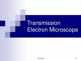
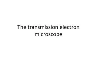
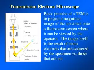
Magnifying using the Electron Microscope © 2007 Paul Billiet ODWS
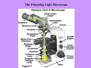
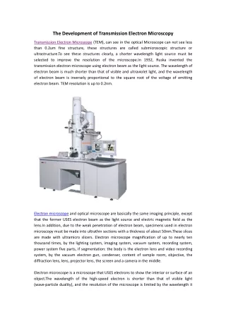
FEATURE LIGHT MICROSCOPE ELECTRON MICROSCOPE Electromagnetic spectrum used Visible light 760nm (red) – 390nm Colours visible Electrons app. 4nm Monochrome Maximum resolving power app. 200nm 0.2nm Fine detail Maximum magnification x1000 – x1500 x500 000 Radiation source Tungsten or quartz halogen lamp High voltage (50kV) tungsten lamp Lenses Glass Magnets Interior Air-filled Vacuum Focussing screen Human eye (retina), photographic film fluorescent (TV) screen, photographic film THE LIGHT MICROSCOPE v THE ELECTRON MICROSCOPE © 2007 Paul Billiet ODWS
FEATURE LIGHT MICROSCOPE ELECTRON MICROSCOPE Preparation of specimens Temporary mounts living or dead Tissues must be dehydrated = dead Fixation Alcohol OsO4 or KMnO4 Embedding Wax Resin Sectioning Hand or microtome slices 20 000nm Whole cells visible Microtome only. Slices 50nm Parts of cells visible Stains Water soluble dyes Heavy metals Support Glass slide Copper grid THE LIGHT MICROSCOPE v THE ELECTRON MICROSCOPE © 2007 Paul Billiet ODWS
Embedding in resin © 2007 Paul Billiet ODWS
Microtome knife © 2007 Paul Billiet ODWS
Copper grid slides © 2007 Paul Billiet ODWS
© 2007 Paul Billiet ODWS CELL STRUCTURE AND MICROSCOPY
CELL STRUCTURE AND MICROSCOPY © 2007 Paul Billiet ODWS