Download
1 / 27
300 likes | 1.44k Vues
Normocytic anemia Fanconi anemia Diamond-Blackfan (PRCA) Types of hemolysis Evidence of hemolysis Reticulocyte G6PD deficiency. Heinz bodies PK deficiency Cytoskeleton Spherocytosis Elliptocytosis PNH. Week 2: Hemolytic Anemia. Normocytic Anemia. Acute hemorrhage Hypo-proliferative

E N D
Normocytic anemia Fanconi anemia Diamond-Blackfan (PRCA) Types of hemolysis Evidence of hemolysis Reticulocyte G6PD deficiency Heinz bodies PK deficiency Cytoskeleton Spherocytosis Elliptocytosis PNH Week 2: Hemolytic Anemia
Normocytic Anemia • Acute hemorrhage • Hypo-proliferative • Acquired aplastic anemia • Constitutional aplastic anemia • Pure red cell aplasia (PRCA)
Acute Hemorrhage • No changes in CBC until plasma volume increases • Later, reticulocytosis
Hypoproliferative Anemia • Pancytopenia • < 25% BM cellularity • Low retic count • Bone marrow defect • Immunologic suppression • Growth Factor deficiency • Abnormal stem cells
Acquired Aplastic Anemia • Idiopathic • Drugs and chemicals (eg, chloramphenicol, benzene, insecticides) • Radiation • Virus • Paroxysmal nocturnal hemoglobinuria
Constitutional Aplastic Anemia • Fanconi’s anemia • Chromosomal breaks
Pure Red Cell Aplasia PRCA • Infections • Drugs • Diamond-Blackfan syndrome • No EPO deficiency
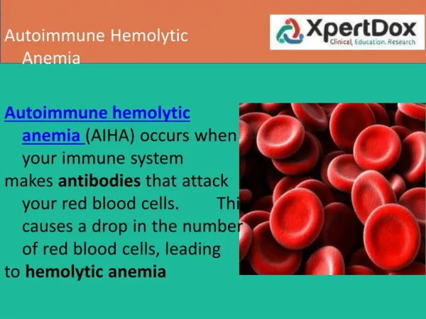
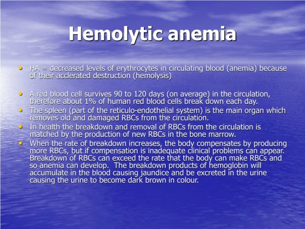
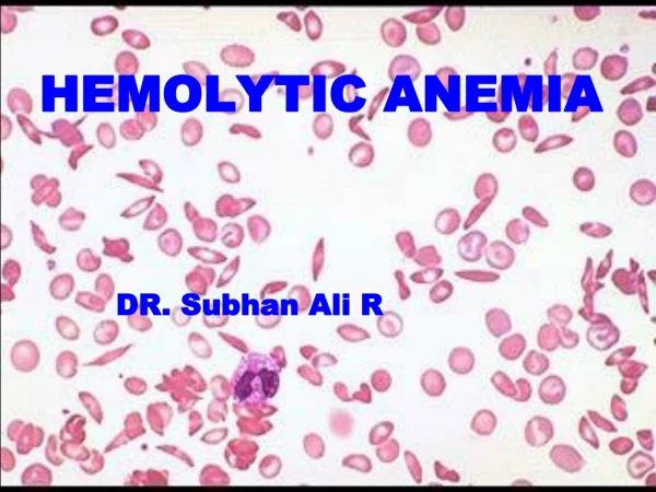
Hemolytic Anemia • Intravascular vs extravascular • Bilirubin metabolism • Types • Membrane defect • Metabolic defect • Extracorpuscular
Evidence of Hemolysis • Low RBC survival with chromium tagging study • Unconjugated bilirubin • Plasma Hb • Decreased serum haptoglobin
Evidence of Erythropoiesis • Polychromasia • Increased reticulocyte • “Shift” macrocytosis • Hypercelluar BM
Correcting Retic Count Retic Index = Retic % x Patient Hct Normal Hct Absolute Retic = Retic % x RBC/mm3 Retic Production Index = Retic Index Days in circulation
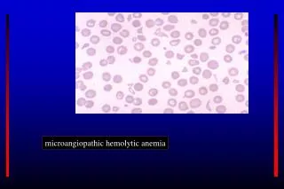
Membrane Defect • Spherocytosis • Elliptocytosis • PNH (sensitivity to complement lysis -- sugar water test, Ham’s test) • Stomatocytosis (possibly Rh null)
Spherocytes Elliptocytes
Paroxysmal Nocturnal Hemoglobinuria • Hematopoietic stem cell disorder • Mutation of phosphatidylinositol glycan class A (PIG-A) gene • Glycosylphosphatidylinositol (GPI) anchors membrane proteins • Without GPI, unable to regulate completment activities on membrane • Hemolysis is pH dependent • Thrombosis can occur
Lab Tests for PNH • Acidified serum lysis test (Ham’s test): PNH cells lyse due to complement activation in acidified serm • Sugar water (sucrose hemolysis) test: RBCs sensitive to complement will lyse in sucrose and serum • Flow cytometry: lack of CD59 on RBCs, or lack of CD59 or CD55 on granulocytes
G6PD deficiency Hexose monophosphate shunt Most common RBC enzyme defect, >50 variants X-linked Low glutathione due to low NADPH Oxidative lysis, Heinz bodies, spherocytic Primaquine, fava beans Pyruvate kinase deficiency Glycolysis Low RBC ATP level Non-spherocytic B12 and folate deficiency Macrocytic HJ bodies Hemoglobinopathies Poikilocytosis Abnormal Hb Metabolic Defect
Heinz body preparation with Crystal violet Unstable hemoglobin
Antibodies Autoimmune Isoimmune Drugs, antibiotics Fresh water Abnormal plasma lipids Acanthocytosis Venom Snake Spider Bee Extracorpuscular Factors
Trauma DIC Hemolytic uremic syndrome (HUS) TTP Angiopathy Heat Heart valves “March” hemoglobinuira Microorganisms Malaria Babesia Clostridium Gram negative endotoxin Extracorpuscular Factors
Schistocytes Malaria