Download
1 / 8
90 likes | 128 Vues
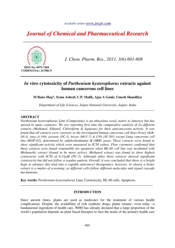
In vitro cytotoxicity of Parthenium hysterophorus extracts against<br>human cancerous cell lines

E N D
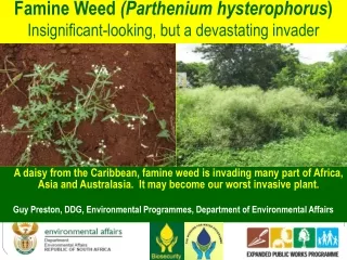
Available online www.jocpr.com Journal of Chemical and Pharmaceutical Research __________________________________________________ J. Chem. Pharm. Res., 2011, 3(6):601-608 ISSN No: 0975-7384 CODEN(USA): JCPRC5 In vitro cytotoxicity of Parthenium hysterophorus extracts against human cancerous cell lines M Raies Haq*, Syma Ashraf, C.P. Malik, Ajaz A Ganie, Umesh Shandilya Department of Life Sciences, Jaipur National University, Jaipur, India ______________________________________________________________________________ ABSTRACT Parthenium hysterophorus Linn (Compositae) is an obnoxious weed, native to America but has spread in many countries. We are reporting first time the comparative analysis of its different extracts (Methanol, Ethanol, Chloroform & Aqueous) for their anticancerous activity. It was found that all extracts were cytotoxic to the investigated human cancerous cell lines Ovary (IGR- OV-I), lung (A-549), prostate (PC-3), breast (MCF-7) & CNS (SF-295) except Lung cancerous cell line(HOP-62), determined by sulphorhodamine B (SRB) assay. These extracts were found to show significant activity which were measured in IC50 values. Flow cytometry confirmed that these extracts were found responsible for apoptosis when HL-60 cell line was incubated with Methanolic extract (found to be more active). Methanol extract was found to show highest cytotoxicity with IC50 of 0.11µM (PC-3). Although other three extracts showed significant cytotoxicity but did not follow a regular pattern. Overall, it was concluded that there is a bright hope to advance this lead into a capable anticancer therapeutics, however, to choose a better extract is a matter of screening, as different cells follow different molecular and signal cascade mechanisms. Key words: Parthenium hysterophorus Linn, Cytotoxicity, HL-60 cells, Apoptosis. _____________________________________________________________________________ INTRODUCTION Since ancient times, plants are used as medicines for the treatment of various health complications. Despite, the availability of rich synthetic drugs, plants remain– even today –a fundamental ingredient of health care. WHO has already declared that a large proportion of the world’s population depends on plant based therapies to face the needs of the primary health care 601
M Raies Haq et al ______________________________________________________________________________ J. Chem. Pharm. Res., 2011, 3(6):601-608 (WHO 1999) [1], P. hysterophorus being one of such medicinal plant, actually an obnoxious weed [2], native to America [3] but has spread in many countries. It is commonly known as Demoina weed (South Africa), parthenium weed (Australia), ragweed Parthenium (USA) and carrot weed or ‘‘Scourge of India’’ or congress weed (India). This alien weed is believed to have been introduced into India as contaminants in PL 480 wheat, imported from the USA in the 1950s. Presently, this invasive weed is widely prevalent in India [4]. It normally grows to a height of about 1 to 1.5m. Chemically complex sesquiterpenoids from this plant spread in air and creates contact dermatitis, direct or indirect sensitization. It has been found to sensitize as many as 56% of occupationally exposed persons without causing apparent dermatitis [5]. Its leaf extracts were found to have a role in the fecundity, fertility and behavioral response [6]. It is the toxic property of this plant that has been exploited to use it as a remedy against various diseases. Its extract is used as folk remedy against skin diseases, ulcerated sores, facial neuralgia, fever and anemia anti-inflammatory[7], shows trypanocidal activity [8], decreases neurotransmitter levels in mice [9], exhibits clastogenic effects, used as a tonic, analgesic, antipyretic, antiperiodic, febrifuge and emmenagogue in Mauritius, Rodrigues, Mexico, Belize and India [10] used as ethno medicine in the treatment of female reproductive problems [11], its allelopathic effects on plants have also been reported [12]. Since, P. hysterophorus and cancer both grow at a rapid speed in many parts of the world, it is important to explore their antagonistic property. So, keeping the implication under contemplation, present study was undertaken to screen the extracts of parthenium for their anticancer activity. EXPERIMENTAL SECTION Chemicals and reagents P. hysterophorus extracts were taken from, Pharmacy Department, Barkatullah university, Bhopal. Human cancer cell lines, breast (MCF-7), ovary (IGR-OV-1), lung (HOP-62), prostate (PC-3), lung (A-549) and CNS (SF-295) and HL-60 were procured from National Centre for cell science (NCCS) (Pune, India). Palcitaxel (taxol), Mitomycin C, Sulphorhodamine B, Streptomycin, Gentamycin, Penicillin and trypsin were purchased from Sigma (St. Louis, MO, USA). Preparation of working solutions of parthenium extracts Four extracts (Aqueous, Ethanol, Methanol, Chloroform) were prepared by dissolving Aqueous extract in10 mg in 1 ml of sterile PBS, whereas, other three extracts were prepared by dissolving 10 mg in 1 ml of DMSO and then these solutions were diluted 10 times (final concentration 1 µg/µl) Preparation of cell suspension for the assay The desired human cancer cell lines were grown at 37oC, 5% CO2 and 90% relative humidity till sub-confluent stage. The cells were harvested by treatment of trypsin- EDTA. The numbers of cells were counted with haemocytometer and cell density was adjusted to 1×10⁶cells /100µl of the cell suspension. Cytotoxicity assay by Sulphorhodamine B dye Before use, 100µl of cell suspension of optimum density was introduced into each well of 96- well plates and incubated for 24h, 37oC, 5% CO2 and 90% relative humidity. 100µl of each extract was added in culture medium to the wells containing the cells. In control wells only 100µl medium was added. The cells with the samples were incubated for 48hours, fixed with ice- 602
M Raies Haq et al ______________________________________________________________________________ J. Chem. Pharm. Res., 2011, 3(6):601-608 cold TCA for 1hour at 4oC. 100µl of SRB was added to each well, staining was done at room temperature for 30 minutes. The SRB solution was removed by washing the plates quickly with water and then 15v/v acetic acid. The bound SRB was solubilised by adding 100µl of 10mM unbuffered tris-Base and absorbances were taken at 540nm. Flow cytometric analysis Effect of Methanolic extract (ME) on DNA content was estimated by cell cycle phase distribution in HL-60 cells. HL- 60 cells (1×10⁶/1.5 ml/well) in 12-well plate were incubated with ME at 25, 50, 75 & 100 µg/ml and staurosporine at 5 µg for 24 h, washed twice with ice- cold PBS, harvested, fixed with ice-cold 70% ethanol, and stored at −20 ◦C for 30 min. After fixation, the cells were incubated with RNase A (0.1 mg/ml) at 37 ◦C for 30 min, stained with propidium iodide (50µg/ml) for 30 min in dark [13]. Cells were analyzed immediately on a SLR flow-cytometer (Becton Dickinson, USA). The fluorescence intensity of sub-G0 cell fraction represents the apoptotic cell population Statistical Analysis All the experiments were carried in triplicate and results were expressed as Mean± SD and statistical analysis of data was done using one way analysis of variance (ANOVA using Tukey- Kramer post-hoc test using PRISM 3 statistical analysis software. RESULTS AND DISCUSSION Effect of p. hysterophorus extracts on percentage growth inhibition SRB assay was used to investigate the potential cytotoxicity effects of Parthenium extracts on six human cell lines. Mito-C and Adriamycin were used as positive controls in this study. The cytotoxicity was presented in percentage growth inhibition of cells as in Table 1. The present study shows various levels of in vitrocytotoxic activities of the Ethanol, Aqueous, Methanolic and Chloroform extracts of p. hysterophorus. The observations from the table 1. showed all extracts are active against cancer cell lines but acted differently. Table 1: percent growth inhibition of different cell lines having different extracts of p. hysterophorus with respect to the control, the concentration of the extracts is 1 µg/µl against different cell lines. Data is in Mean ± S.D and representative of three similar experiments. This implies that the active extracts have specific cytotoxic activities against specific cell lines. The activity was shown using 100µl of extracts (1µg/µl). The Methanol extract was found to show highest percentage of growth inhibition with all the five cell lines that ranged from 78- 98%, Chloroform extract showed growth inhibition in the range of 62% (prostate-PC-3) to 94% Parthenium extracts Breast Ovary IGR-OV-1 HOP-62 percentage growth inhibition 94 ± 1.36 0 87 ± 1.44 0 98 ± 1.14 8 ± 0.38 81 ± 0.89 78 ± 5.58 93 ± 0.58 92 ± 4.00 0 71 ± 7.48 72 ± 1.89 71 ± 3.68 - - 80 ± 5.90 - - - Lung Prostate PC-3 Lung A-549 CNS SF-295 MCF-7 Chloroform Ethanol Methanol Aqueous Mito-C Adriamycin 93 ± 0.68 77 ± 0.30 93 ± 2.17 92 ± 4.00 - 86 ± 6.87 62 ± 0.64 68 ± 0.80 73 ± 0.48 56 ± 0.74 57 ± 1.29 56 ± 1.13 - - - 85 ± 5.09 603
M Raies Haq et al ______________________________________________________________________________ J. Chem. Pharm. Res., 2011, 3(6):601-608 (ovary-IGR-OV-1) in five cell lines, Ethanolic extract showed growth inhibition in the range of 56% Prostate (PC-3)& CNS (SF-295) to 87% Ovary (IGR-OV-1) in five cell lines, Aqueous extract showed growth inhibition in the range of 71 %( prostate-PC-3) & CNS (SF-295) to 92% (ovary-IGR- OV-1) & breast (MCF-7) in five cell lines- indicating their anticancer activity but no extract showed was effective against lung (HOP-62) cell line as percentage growth inhibition was only 0-8%. Lung (HOP-62) being unresponsive so it was not shown in graphical presentation for the sake of simplicity as shown in figure 1. Results as shown in table 2 indicated that Methanol extract has highest activity that ranged from 0.11- 0.45. SF-295 has lesser activity (0.51) with Ethanol extract while as other three extracts have around same activity (0.45, 0.46 &0.47) with this cell line. All the four extracts were found to have almost same activity with MCF-7 cell line. PC-3 has highest activity (0.11) with Methanol extract, lowest with Ethanol extract (0.39) and around same activity (0.27& 0.29) with Chloroform and Aqueous. A-549 has highest activity (0.17) with Methanol and lowest with Ethanol extract (0.46). IG-OV-1 has highest activity with Aqueous extract (0.16) and lowest with Ethanol (0.25) as shown in figure 2. Fig 1: In vitro cytotoxicty of P. hysterophorus extracts on various cell lines. Data is represented as Mean±SD. (Mito-c = Mitomycin C, CE= Chloroform extract, EE= Ethanol extract, ME= Methanol extract, AE= Aqueous extract) Table 2: IC50 Values (µM) of standard Cyclophosphamide Monohydrate and four different extracts of P. hysterophorus against Human Cancerous Cell Lines IGR-OV-I, A-549, PC-3, MCF-7& SF-295. Substance Cyclophosphamide monohydrate 0.09 ± 0.02 0.11 ± 0.01 0.07 ± 0.01 0.22 ± 0.03 0.20 ± 0.02 Chloroform 0.21 ± 0.03 0.34 ± 0.06 0.27 ± 0.03 0.47 ± 0.08 0.46 ± 0.06 Ethanol 0.25 ± 0.04 0.46 ± 0.07 0.39 ± 0.05 0.48 ± 0.07 0.51 ± 0.06 Methanol 0.19 ±0.03 0.17 ±0.02 Aqueous 0.16 ±0.02 0.21 ±0.04 Human Cancerous Cell lines PC-3 IGR-OV-1 A-549 MCF-7 SF-295 0.11 ± 0.01 0.45 ± 0.05 0.29 ± 0.04 0.46 ± 0.09 0.45 ± 0.02 0.47 ± 0.06 The cells were grown in 96-well culture plate were treated with four extracts for 48 h, thereafter cultures incubated with SRB. Data in in Mean ± S.D and representative of three similar experiments. 604
M Raies Haq et al ______________________________________________________________________________ J. Chem. Pharm. Res., 2011, 3(6):601-608 Fig 2: Showing IC50 valves of different extracts and control cyclophosphamide monohydrate. Data is represented as Mean ± SD. (CM=cyclophosphamide Monohydrate, CE= Chloroform Extract, EE= Ethanol Extract, ME= Methanol Extract, AE= Aqueous Extract) Methanol extract (ME) increases sub-G0 DNA fraction of cell cycle phase distribution Since Methanol extract was found to show highest percentage growth inhibition and activity with the human cancerous cell lines, so, it was selected for determination of apoptosis in HL-60 cell lines by flow cytometry. Effect of ME on DNA content by cell cycle phase distribution was assessed using human leukemia HL-60 cells. ME exhibited concentration dependent increase in hypo diploid sub-G0 DNA fraction (<2n DNA) (Fig.3). The sub-G0 fraction was 8.5% in control cells, which increased to∼47.4% at 100µg/ml of ME treatment. Further, the cell cycle G2/M phase was not affected indicating that ME does not produce any mitotic block or caused delay in cell cycle as shown in figure 4. Since anticancer drugs have minimum side effects, so to select a drug having a particular specificity towards a target cell line will be appreciated [14]. Keeping the above need under consideration, four extracts (Aqueous, Chloroform, Methanol and Ethanol) of P. hysterophorus were evaluated for their cytotoxic effect on human cancerous cell lines in order to select the effective extract for all cell line lines and most important being that, it was the first attempt to reveal the operative comparative analysis of different extracts of P. hysterophorus on human cancerous cell lines to evaluate the anticancerous activity, but it was found that all the four extracts showed different reactivity towards different cell lines. Methanol extract was found to show both highest percentage growth inhibition and activity (IC50 values), although other three extracts also showed significant cytotoxicity but did not follow a regular pattern, so, it is difficult to select a particular extract for anticancerous activity and the selection is a matter of screening as solubility of diverse compounds is different in various solvents or different molecular and cascade mechanisms followed by different cell lines, however, the exact mechanism is not known. 605
M Raies Haq et al ______________________________________________________________________________ J. Chem. Pharm. Res., 2011, 3(6):601-608 Staurosporine, 12.5µg/ml 49.36% FL2-A 8.5% ME ME 75µg/ml 35.9% 100µg/ml 47.4% ME ME 25µg/ml 10.9% 50µg/ml 18.2% Fig 3: DNA content analyses in Methanol Extract (ME) treated HL-60 cells. Cells (1×10⁶/1.5 ml/12-well plates) were exposed to indicate concentrations of me for 24 h and stained with PI to determine DNA fraction of cells for hypo-diploid (sub-G0,<2n DNA) population analyzed from Fl2-A vs. cell counts is shown (%). data are representative of one of three similar experiments Fig 4: Flow Cytometric analysis of ME-induced apoptosis in HL-60 cells. Cells (1×10⁶/1.5 ml/12-well plates) were incubated with different concentrations of ME for 24 h to analyze apoptosis. Data is presented as Mean ± SD. Data are representative of one of three similar experiments. (ME = Methanol EXTRACT, ME1 = 25µg/ml, ME2 = 50µg/ml, ME3=75µg/ml, ME 4= 100µg/ml and ST= Staurosporine 606
M Raies Haq et al ______________________________________________________________________________ J. Chem. Pharm. Res., 2011, 3(6):601-608 Present study revealed that all the extracts were cytotoxic to five human cancerous cell lines (IGR-OV-1, A-549, PC-3, MCF-7, & SF-295) but not for lung cancerous cell line(HOP-6), our results were in accordance with Das et al. [15] where it was found that acetylated pseudoguaianolides from P. hysterophorus had cytotoxicity against human T lymphocyte, HL-60 and Hela cell lines. Further, our results show that breast( MCF-7) and ovary (IGR-OV-1) cell lines showed a maximum responses with all the four extracts where percentage growth inhibition was in the range of 77-98% as shown in table 1 however there was not a regular pattern. Although other three cell lines also showed a significant growth inhibition but it was found less effective than former (cell growth inhibition 56-93%). Moreover, it was found that Methanol extract showing higher activity (IC50 = 0.11-0.45µM) with all the five cell lines as shown in table 2. Taking into account the prospective of P. hysterophorus extracts as cytotoxic/anticancerous agents, we advanced our study to understand the mechanism of apoptotic cell death. Apoptosis is a foremost form of cell death, characterized by a chain of stereotypic morphological changes such as formation of apoptotic bodies, chromatin condensation, shrinkage of cells and bleb formation, internuclesomal DNA fragmentation and sub-G0 DNA accumulation [16]. DNA fragmentation is a relatively late event and hallmark of apoptosis. To confirm apoptosis induced by Methanol extract, having higher activity as shown in table 2 , we applied flow cytometry to find out the apoptosis, Methanol extract significantly induce the apoptotic body’s formation and increase in hypo diploid sub-G0 DNA fraction (<2nDNA) of HL-60 cells. Overall, it was found that all extracts of the p.hyterophus have cytotoxic effects on human cancerous cell lines studied, but the best extract showing uniform reactivity was Methanol and other three extracts did not follow a regular pattern. IC50 values supported the view and flow cytometry confirmed the apoptotic nature of these extracts through measurement of fluorescence. CONCLUSION The present studies lead us to conclude that all P. hysterophorus extracts exhibited cytotoxic effect on wide range of human cancer cell lines and to select a better extract is a matter of screening. Methanol extract showed highest percentage growth inhibition and activity and other three extracts did not follow a regular pattern because of different cell specificity and cascade mechanisms followed by different cells. Further, these extracts have a role in apoptosis when analysed in human leukemia HL-60 cells. In future there is a bright hope to advance this lead into a capable anticancer therapeutics. Acknowledgements We wish to thank Jaipur National University, Jaipur for granting funds that made this work successful. We also thank Indian Institute of Integrative Medicine, Jammu for their help for performing Fow Cytometry. We acknowledge the excellent assistance of Meena and Divya for discussions and critical comments about the manuscript. REFERENCES [1] Dikshit A., Shahi S. K., Pandey K. P., Patra M. and Shukla A. C., Nat. Acad. Sci. Letters., 2004, 27 [5&6)], 145-164. [2] Towers G. H. N., Mitchel J.C., Rodriguez E., Bannet D. F. and Subba P., J. Sci. Ind. Res., 1977, 36, 672–684. 607
M Raies Haq et al ______________________________________________________________________________ J. Chem. Pharm. Res., 2011, 3(6):601-608 [3] Navie S. C., McFadyen R. E., Panetta F. D. and Adkins S. W., Plant Protection Quarterly.,1996, 11, 76-88. [4] Shah B. A., Kaur R. and Gupta P., et al, Biorg. Med. Chem. Lett., 2009, 19[15], 4394- 4398. [5] Rao P. V., Mangala A., Rao B. S. and Parkash K. M., Experientia., 1977, 33: 1387. [6] Kumar S., Singh A. P. and Nair G., et al, Parasitol. Res., 2011, 108 [4], 853-859. [7] Recio M. C., Giner R. M. and Uriburu L., et al, Life Sci., 2000, 66 [26], 2509- 2518. [8] Talakal T. S., Dwivedi S. K., Sharma S. R., Indian J. Ex. Biol., 1995, 33, 894-896. [9] Verma S., Shrivastava R., Prasad P. K. and Shrivastava V. K., Phytother Res., 2007, 21, 183-185. [10] Hirschhorn H., J Ethnopharmacol., 1981, 4[2], 129-158. [11] Ramos A., Rivero R., Visozo A., Piloto J. and Garcia A., Mutat. Res.,2002, 514: 19-27. [12] Patil T. M., Hedge B. A., Curr. Sci.,1988, 42, 1178-1181. [13] Kidd V. J., Annu. Rev. Physiol.,1988, 60, 533–573. [14] Skehan P., Storeng R. and Scudiero D., J. Natl. Cancer Inst., 1990, 82[13], 1107-12. [15] Das B., Reddy V. S. and Krishnaiah M., Phytochemistry., 2007, 68[15], 2029-2034. [16] Bhushan S., Singh J., Rao J. M., Saxena A .K. and Qazi G.N., Nitric Oxide.,2006, 14, 72–88. 608