Download
1 / 8
191 likes | 556 Vues
Anatomy and physiology of the eye makes it a highly protected organ. Designing an effective therapy for ocular diseases, especially for the posterior segment, has been considered as aformidable task. Limitations of conventional route of administration have challenged scientists to find alternative mode of administration like periocular routes. Targeted drug delivery hasgenerated a great deal of interest in the field because of its potential to overcome many barriers associated with current therapy. Application of nanotechnology has been very promising in the treatment of a gamut of diseases. In this review, we have briefly discussed several novel ocular drug delivery systems such as microemulsions, nanosuspensions, nanoparticles, liposomes, niosomes, dendrimers, implants, and hydrogels. Current momentum in the invention of new drug delivery systems hold a promise towards much improved therapies for the treatment of vision threatening disorders.

E N D
Available on line www.jocpr.com Journal of Chemical and Pharmaceutical Research __________________________________________________ J. Chem. Pharm. Res., 2010, 2(3):348-355 ISSN No: 0975-7384 CODEN(USA): JCPRC5 Novel ocular drug delivery systems: An overview Akanksha Tiwari1* and Raj Kumar Shukla2 1Baroda College of Pharmacy, Parul Trust, Limbda, Waghodia, Vadodara, Gujrat 2R&D –NDDS Department, Sun Pharma Industries Ltd., Sun Pharma Advance Research Centre, Tandalja Vadodara, Gujrat, India ______________________________________________________________________________ ABSTRACT Anatomy and physiology of the eye makes it a highly protected organ. Designing an effective therapy for ocular diseases, especially for the posterior segment, has been considered as a formidable task. Limitations of conventional route of administration have challenged scientists to find alternative mode of administration like periocular routes. Targeted drug delivery has generated a great deal of interest in the field because of its potential to overcome many barriers associated with current therapy. Application of nanotechnology has been very promising in the treatment of a gamut of diseases. In this review, we have briefly discussed several novel ocular drug delivery systems such as microemulsions, nanosuspensions, nanoparticles, liposomes, niosomes, dendrimers, implants, and hydrogels. Current momentum in the invention of new drug delivery systems hold a promise towards much improved therapies for the treatment of vision threatening disorders. Key words: Nanotechnology, Targeted drug delivery, Enhanced Bioavailability. _____________________________________________________________________________ INTRODUCTION Ocular drug delivery has remained as one of the most challenging task for pharmaceutical scientists. The unique structure of the eye restricts the entry of drug molecules at the required site of action. Drug delivery to the eye can be broadly classified into anterior and posterior segments. 348
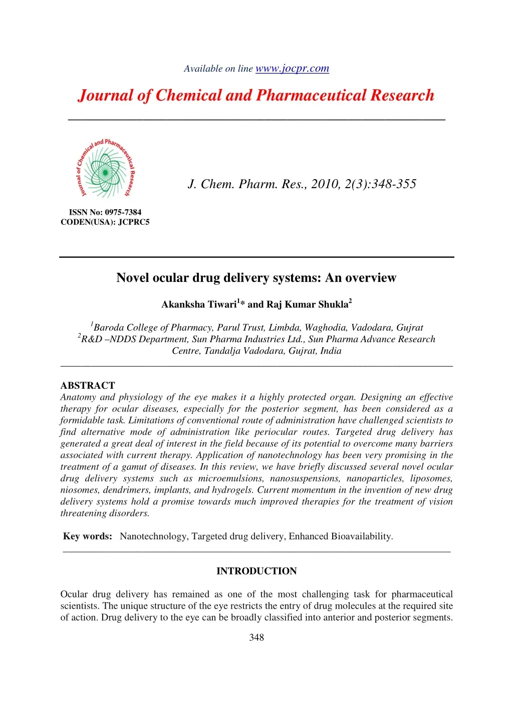
Akanksha Tiwariet al J. Chem. Pharm. Res., 2010, 2(3):348-355 _____________________________________________________________________________ Conventional systems like eye drops, suspensions and ointments cannot be considered optimal in the treatment of vision threatening ocular diseases [1]. However, more than 90% of the marketed ophthalmic formulations are in the form of eye drops. These formulations mainly target the anterior segment eye diseases [2]. Most of the topically applied drugs are washed off from the eye by various mechanisms (lacrimation, tear dilution and tear turnover) resulting in low ocular bioavailability of drugs. Moreover, human cornea comprising of epithelium, substantia propria and endothelium also restricts the ocular entry of drug molecules [3]. As a result of these factors less than 5% of administered drug enters the eye. Alternative approaches like incorporation of permeation enhancers/cyclodextrins and increasing the viscosity of solutions did not provide any significant improvement. Recently many drug efflux pumps have been identified and significant enhancement in ocular drug absorption was achieved following their inhibition or evasion. But prolonged use of such inhibitors may result in undesirable effects [4]. Treatment of posterior segment diseases still remains a herculean task for the formulation scientists. The tight junctions of blood retinal barrier (BRB) restrict the entry of systemically administered drugs into the retina [5]. High vitreal drug concentrations are required in the treatment of posterior segment diseases. This can be made possible only with the local administration (intravitreal injections/implants and periocular injections). Periocular injections are associated with fairly high patient compliance as compared to intravitreal injections [6]. Dramatic changes have been observed in the field of ocular drug delivery over a decade. Insight into various membrane transporters/receptors present on the eye opened a new window of opportunities. Especially polar drug molecules, which fail to permeate ocular barriers, can be conveniently delivered via targeted drug delivery systems [7]. Overview on anatomy and diseases affecting eye Broadly we discuss the structure of eye under two subheadings (a) anterior segment and (b) posterior segment.Anterior segment consists of front one-third of eye that mainly includes pupil, cornea, iris, ciliary body, aqueous humor, and lens while the posterior segment consists of the back two-thirds of the eye that includes vitreous humor, retina, choroid, macula, and optic nerve (Fig. 1). [8], [9] Figure 1: Structure of Eye Novel ocular drug delivery systems Colloidal carriers have been widely exploited in the field of drug delivery science. It provides a more selective targeting along with sustained release of molecules at the desired site. Applications of nanotechnology can be very exciting in the treatment of a gamut of diseases affecting the anterior as well as the posterior segment of the eye. An ideal therapy requires 349
Akanksha Tiwariet al J. Chem. Pharm. Res., 2010, 2(3):348-355 _____________________________________________________________________________ selectively targeting of active agent to various diseases like CNV, diabetic retinopathy and solid tumors in the eye. Retina does not possess lymphatic system moreover angiogenesis in this part of eye has similar features to the solid tumor with enhanced permeability and retention (EPR) effects [10]. Delivery of a drug via nanotechnology based product fulfills mainly three objectives as follows (a) enhances drug permeation (b) controls the release of drug (c) targets drug [11]. Enhancement in bioavailability Topical bioavailability can be improved by maximizing precorneal drug absorption and minimizing precorneal drug loss. Viscosity improver: The viscosity enhancers used are hydrophilic polymers such as cellulose, polyalcohol and polyacrylic acid. Sodium carboxy methyl cellulose is one of the most important mucoadhesion polymers having mono adhesive strength [12]. The effects of polyacrylic acid and polyacrylamide based hydrogels are tested on miotic response of pilocarpine. Carbomer were used in liquid and semisolid formulations as suspending or viscosity increasing agents. Hyaluronic acid offers a biocompatible and biodegradable matrix for fabrication of ocular sustained release dosage form- Dosage forms based on the benzyl esters of hyaluronic acid were used for ophthalmic sustained release of methyl prednisolone. Films and microspheres were also prepared from hyaluronic acid. Polysaccharide such as xanthan gum was found to increase the viscosity [13]. Viscosity vehicles increases the contact time and no marked sustaining effect are seen. Penetration enhancers: They act by increasing corneal uptake by modifying the integrity of corneal epithelium. Chelating agents, preservatives, surfactants and bile salts were studied as possible penetration enhances [14]. Prodrugs: Prodrugs enhance comeal drug permeability through modification of the hydrophilic or lipophilicity of the drug .[15] The method includes modification of chemical structure of the drug molecule, thus making it selective, site specific and a safe ocular drug delivery system. Drugs with increased penetrability through prodrug formulations are-epinehrine1, phenylephrine, timolol, pilocarpine and albuterol [16]. Bioadhesive polymers: These polymers [17] adhere to the mucin coat covering the conjunctiva and the corneal surfaces of the eye, thus prolonging the residence time of a drug in the conjunctival sac. These polymers can be neutral, synthetic or semi synthetic. Polyacrylic acid, polycarbophil and hyaluronic acid are synthetic polymers commonly used. Chitosan is a bioadhesive vehicle suitable for ophthalmic formulation. Xanthan and carrageenan are also described as bioadhesive polysaccharides [18]. 350
Akanksha Tiwariet al J. Chem. Pharm. Res., 2010, 2(3):348-355 _____________________________________________________________________________ Enhancement in controlled drug-delivery It is realized that the preferred system of ophthalmic delivery would provide improved bioavailability, site-specific delivery and with continuous drug release. So achievements have been made in the following areas: In situ forming gels: The progress has been made in gel technology lip the development of droppiable gel. They are liquid upon instillation and undergo phase transition in the ocular cul-de-sac to form visco-elastic gel and this provides a response to environmental changes [19]. Oil in water emulsions: Phospholipids and pluronics were used as the emulsifiers. Antioxidants were added to improve their shelf-life. The intra-ocular pressure reducing effect of a single, topically administered dose of a pilocarpine emulsion lasted for 29 h in rabbits compared to generic pilocarpine solution which lasted only for 5hr [20]. Oil in water emulsion is useful for delivery of Water insoluble drugs, which is solubilised in the internal oil phase. Colloidal Carriers: Nanoparticles: According to Sahoo et al., “Nanoparticles are defined as particles with a diameter of less than 1 µm, comprising of various biodegradable or non biodegradable polymers, lipids, phospholipids or metals” [21]. They can be classified as nanospheres or nanocapsules depending upon whether the drug has been uniformly dispersed or coated within polymeric material. Nanoparticles provide sustained release-and prolonged therapeutic activity when retained in the cul-de-sac after topical administration and the entrapped drug must be released from the particles at an appropriate rate. Most commonly used polymers are venous poly (alkyl cyanoacrylates), poly S- caprolactone and polylactic-co-glycolic acid, which undergo hydrolysis in tears [22] Chitosan coated poly (epsilon-caprolactone)nanoparticlesof enhancement in ocular bioavailability [23]. Enhanced permeation across the cornea was also observed when poly (epsilon-caprolactone) nanoparticles were coated with polyethylene glycol [24]. Mucoadhesive chitosan-sodium alginate nanoparticles were prepared and evaluated for topical delivery of gatifloxacin.This system resulted in burst release during the first hour followed by sustained release for 24 h. This approach helps in reducing the dosing frequency of the antibiotic because of the sustained action observed after single administration [25]. Niosomes: Niosomeswere first reported in the seventies as a feature of the cosmetic industry by Vanlerberghe et al., [26] Handjani-vila et al., [27] Van Abbe [28] explained that the non – ionic surfactants are preferred because the irritation power of surfactants decreases in the following order: cationic > anionic > ampholytic > non-ionic. Green and Downs [29], Keller et al. [30], Burstein [31], Kaur and Smitha [32] reported that an increased ocular bioavailability of water soluble, entrapped in niosomes, may be due to the fact that surfactants also act as penetration enhancers as they can remove the mucus layer and break functional complexes. Singh and Mezei [33] stated that niosomes are a suitable delivery system for both hydrophilic and lipophilic drugs. Vyas et al. [34] prepared both niosomes and discomes of water-soluble drug timolol maleate and found that discomes entrapped comparatively a higher amount of drug (25% as compared to 14% indomethacin resulted in twofold 351
Akanksha Tiwariet al J. Chem. Pharm. Res., 2010, 2(3):348-355 _____________________________________________________________________________ in case of niosomes). Moreover, an increase in ocular bioavailability was found to be approximately 3.07-fold compared to 2.48-fold in case of niosomes with respect to timolol maleate Ghada abdelbary [35] and Nashwa el-gendy investigated the feasibility of using non- ionic surfactant vesicles as carriers for the ophthalmic controlled delivery of a water soluble local antibiotic, Gentamicin sulphate. Liposomes: Liposomes are lipid vesicles containing aqueous core which have been widely exploited in ocular delivery for various drug molecules.Liposomes are favorable for lipophilic drugs and not for-hydrophilic drugs. liposomes has an affinity to bind to, ocular surfaces, and release contents at optimal rates [36]. Positively charged liposomes have a greater affinity, to increase both precorneal drug retention and drug bioavailability .The addition of stearylamine to a liposomal preparation enhanced the corneal absorption of dexamethyl valerate. The corneal epithelium is thinly coated with negatively charged mucin to which the positive surface charge of the liposome may absorb more strongly. Coating with bioadhesive polymers to liposomes, prolong the precomea retention of liposomes. Carbopol 1342-coated pilocarpine containing liposomes were shown to produce a longer duration of action [37].Ciprofloxacin (CPFX) was also formulated in liposomal environmental which lowered tear-driven dilution in the conjunctival sac. Multilamellar vesicles from lecithin and alpha-L-dipalmithoyl-phosphatidylcholine were used to prepare liposome containing CPFX. This approach produced sustained release of the drug depending on the nature of the lipid composition selected [38]. Microparticulates: They are drug containing, micron sized polymeric particles suspended in a liquid medium. Drugs can be physically dispersed in the polymer backbone [39]. The drug is released in cul-de- sac through diffusion, chemical reaction, and polymer degradation and micro particles are larger than nanoparticles. Acyclovir loaded chitosan microspheres [40] and Pilocarpine-loaded albumin or gelatin microspheres.are available. Microparticulate technology has the advantage of better patient acceptability, since they can be topically administered as an eye drop [41]. Microemulsion: Microemulsions are dispersions of water and oil facilitated by a combination of surfactant and co-surfactant in a manner to reduce interfacial tension. These systems are usually characterized by higher thermodynamic stability, small droplet size (100 nm) and clear appearance [42]. Microemulsion systems have also been exploited to improve permeation across the cornea. An oil in-water system consisting of pilocarpine using lecithin, propylene glycol, PEG 200 as surfactant/co surfactants, and isopropyl myristate as the oil phase has been designed, which is nonirritating to the rabbit animal model [43]. Such formulations often provide sustained drug release thereby reducing frequency of the drug administration. In case of pilocarpine, microemulsion based system lowers the frequency of administration to two times as compared to four times with conventional eye drops in a day. This was due to enhancement of the permeation by surfactant-co-surfactant combination. Nanosuspensions: Nanosuspenisons usually consist of colloidal carriers like polymeric resins which are inert in nature. They help in enhancement of drug solubility and thus bioavailability. Unlike 352
Akanksha Tiwariet al J. Chem. Pharm. Res., 2010, 2(3):348-355 _____________________________________________________________________________ microemulsions, they are also popular because of their non irritant nature. Flurbiprofen encapsulated in eudragit RS 100® and RL 100® polymer resins prevents myosis, which might be induced during extracapsular cataract surgery [44] Animal studies have revealed that anti- inflammatory effect of nanosuspensions was more than microsuspensions. Similar studies were carried out using piroxicam in eudragit RS 100. In vivo studies in rabbits have shown significant anti-inflammatory effects compared to microsuspensions [45]. Dendrimers: Dendrimers are macromolecular compounds made up of a series of branches around a central core. Their nanosize, ease of preparation, functionalization and possibility to attach multiple surface groups render them suitable alternative vehicle for ophthalmic drug delivery” [46]. Iontophoresis: Ocular iontophoresis has gained significant interest recently due to its non-invasive nature of delivery to both anterior and posterior segment. It requires a mild electric current which is applied to enhance ionized drug penetration into tissue. This mode of delivery can overcome the potential side effects associated with intraocular injections and implants mentioned earlier. OcuPhor™ system has been designed with an applicator, dispersive electrode and a dose controller for transscleral iontophoresis (DDT) [47]. CONCLUSION Effective treatment of ocular diseases is a formidable challenge for scientists in the field especially because of the nature of diseases and presence of the ocular barriers especially in posterior ocular segments. An ideal therapy should maintain effective levels of drug for the longer duration following a single application. Drug delivery by topical and intravitreal routes cannot be considered safe, effective and patient friendly. Drug delivery by periocular route can potentially overcome many of these limitations and also can provide sustained drug levels in ocular pathologies affecting both segments. Transporter targeted delivery can be a promising strategy for many drug molecules. Colloidal carriers can substantially improve the current therapy and may emerge as an alternative following their periocular administration. Acknowledgements Authors are thankful to the authorities of Baroda College of Pharmacy for providing necessary facilities and library. REFERENCES [1]D Aggarwal; I P Kaur. Int. J. Pharm.,2005;290, 155. [2]C Bharath; S R Hiremath . Pharmazie, 1999; 51, 55. [3]C L Bourlais; L Acar; H Zia; PA Sado; T Needham; R Leverge Prog. Retin. Eye Res. 1998;17:33– 58 [4]R Jain; S Majumdar; Y. Nashed; D Pal; A K Mitra. Mol. Pharm. 2004; 1:290–299. [5]K G Janoria; S Gunda; S H S Boddu; A K Mitra.Expert Opin Drug Deliv. 2007; 4:371–388. [6]S Raghava; M Hammond; U B Kompella. Expert Opin Drug Deliv. 2004; 1:99–114. [7]C S Dias; B S Anand; A K Mitra. J. Pharm. Sci. 2002; 91:660–668. 353
Akanksha Tiwariet al J. Chem. Pharm. Res., 2010, 2(3):348-355 _____________________________________________________________________________ [8]F Latorre; AP Nicolal. Drugs Exp, Clin, Res.1998; 24, 153. [9]Lee, V.H.L. and Robinson, J.R., J. Ocul. Pharmacol,1986; 2, 67. [10]T Yasukawa; Y Ogura; Y Tabata; H Kimura; P Wiedemann ;Y Honda .Drug delivery systems for vitreoretinal diseases.Prog. Retin. Eye Res.2004; 23:253–281. [11]S K Sahoo; F Dilnawaz; S Krishna Kumar. Drug Discov Today. 2008; 13:144–151. [12]Davios; Davies Clin. Exp, Pharmasol. Physiol., 90W. 27, 558. [13]E L Gazayerly; N Omaima; AN H Hikal. Int. J. Pharm. 1997; 158,121. [14]J C Keistea; E RCooper; P J Missel; J C Long; D F Hager. J. Pharm. Sci, 1991; 80, 50. [15]A J Khopade; N K Jain. Pharmazie, 19%. 50, 812. [16]S Kumar; B O Haglund ; K J Himmelstein. J. Ocul.Pharmcol., 1994; 10, 47. [17]F Latorre; A P Nicolal. Drugs Exp, Clin, Res. 1998; 24, 153. [18]V H L Lee; J R Robinson. J. Ocul. Pharmacol., 1986, 2, 67. [19]H R Lin; K C Sung. J. Contol. release, 2000; 69, 379. [20]G Meseuger; R Gumy; P Buri; A Rozier; B Plazonnet. Int. J. Pharm., 1993; 95, 229. [21]S K Sahoo; F. Dilnawaz; S Krishnakumar. Drug Discov Today. 2008; 13:144–151. [22]K S Rathore ; R K Nema . International Journal of Pharm tech Research, 2009; Vol.1, No.2, pp 164-169. [23]P Calvo; J L Vila-Jato ; M J Alonso. J Pharm Sci. 1996; 85:530–536. [24]AM De Campos; A Sanchez; R Gref; P Calvo; and M J Alonso. Eur. J. Pharm. Sci. 2003; 20:73– 81. [25]S K Motwani ; S Chopra ; S Talegaonkar ; K Kohli; F J Ahmad ;R.K. Khar Eur.J.Pharm .Biopharm.2008; 68:513–525. [26]G Vanderburgh; R M Handjani-Vila; C Berthelot ;H Sebag . Carl Hanser Verlag, Zurich. 1972. [27]R M Handjani-Vila ; A Rlbier ;B Rondot ; G Vanlerberghe . Int. J. Cosmetic Sci.1979; 1, 303-314. [28]N J Van Abbe . J Soc Cosmet. Chem. 1973; 24, 685-687. [29]K Green; S , Arch.Ophthalmol.1975; 93, 1165- 1168. [30]N Keller; D Moore; D Carper Longiwell A. Exp. Eye.Res. 1980; 30, 203-210. [31]Burstein NL. Invest. Ophthalmol. Vis. Sci. 1984; 25, 1453. [32]I P Kaur; R Smitha., Drug Dev. Ind. Pharm.2002;28, 353-369 [33]K Singh; M Mezei . Int. J. Pharm. 1984;19, 263-269. [34]S P Vyas; N Mysore ; V Jaittley ; N Venkatesan . Pharmazie 1998; 53,466-469. [35]Ghada abdelbary, Nashwa el-gendy, , AAPS Pharm.Sci. Tech,2008; 9(3). [36]D L Middleton; S H S Leung; J R Robinson;V Lenaerts R Gummy Eds; Bioadhesive Drug Delivery Systems, CRC Press, Boca Raton. 1990; 203. [37]A S Monem; F M Ali; M W Ismail. Int. J. Pherm., 2000;198, 20. [38]R M Hathout; S Mansour; N D Mortada; A S Guinedi. Liposomes as an ocular delivery system for acetazolamide: in vitro and in vivo studies. AAPS Pharm Sci Tech. 2007,; 8(1). [39]A Urtti; H RouhWnen; T Kaila; V Saano .Pharm.Res.1994;11, 1278. [40]G Wei; H Xu; PT U SM Ding; J M J Zheng. J Control. Release, 2002; 83, 65. [41]Zimmer, A K Chetoni; R Saettone, M P Zerbe; H J Kreuter. J. Control. Release, 1995; 33, 31. [42]M J Ansari; K Kohli; N Dixit. PDA J. Pharm. Sci. Technol. 2008; 62:66–79. [43]Hasse; S Keiper. Eur.J.PharmBiopharm.1997; 43:179– 183. 354
Akanksha Tiwariet al J. Chem. Pharm. Res., 2010, 2(3):348-355 _____________________________________________________________________________ [44]I R Pignatello; C Bucolo; G Spedalieri; A Maltese; G Puglisi. Biomaterials.2002; 23:3247– 3255 [45]K Adibkia; M R Siahi Shadbad; A Nokhodchi; A Javadzedeh; M arzegar- Jalali, J Barar; G Mohammadi; Y Omidi. J. Drug Target.2007; 15:407–416. [46]N Ogata; T Otsuji ; M Matsushima; T Kimoto; R Yamanaka; K Takahashi; M.Wada; M Uyama; Y Kaneda. Curr. Eye Res. 1999; 18:261– 269. [47]TM Parkinson; E Ferguson; S Febbraro; A Bakhtyari ; M King; M Mundasad. J. Ocul. Pharmacol. Ther. 2003;19:145– 151. 355