Download
1 / 19
290 likes | 691 Vues
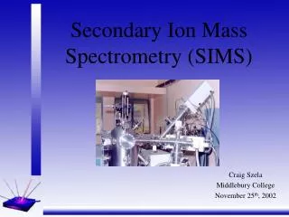
Secondary Ion Mass Spectrometry (SIMS). Advisor:Kuen Shian Wu Student:Kuen Chi Wu. Outline. Introduction Mass Analyser Secondary Ion Detectors Results and Discussion References.

E N D
Secondary Ion Mass Spectrometry(SIMS) Advisor:Kuen Shian Wu Student:Kuen Chi Wu
Outline • Introduction • Mass Analyser • Secondary Ion Detectors • Results and Discussion • References
In 1910 British physicist JJ Thomson observed a release of positive ions and neutral atoms from a solid surface induced by ion bombardment. Improved vacuum pump technology in the 1940s enabled the first prototype experiments on SIMS by Herzog and Viehböck in 1949, at the University of Vienna , Austria. Introduction
SIMS is widely used for analysis of trace elements in solid materials, especially semiconductors and thin films. The SIMS primary ion beam can be focused to less than 1 um in diameter. Controlling where the primary ion beam strikes the sample surface provides for microanalysis, the measurement of the lateral distribution of elements on a microscopic scale. During SIMS analysis, the sample surface is slowly sputtered away. This slow sputtering mode is called static SIMS in contrast to dynamic SIMS used for depth profiles. Introduction
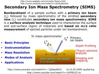
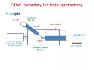
Secondary ion mass spectrometry (SIMS) is based on the observation that charged particles (Secondary Ions) are ejected from a sample surface when bombarded by a primary beam of heavy particles. A basic SIMS instrument will, therefore, consist of: A primary beam source to supply the bombarding species. A target or sample that must be solid and stable in a vacuum. A method of collecting the ejected secondary ions. A mass analyser to isolate the ion of interest . An ion detection system to record the magnitude of the secondary ion signal. Introduction
Introduction In the field of surface analysis, it is usual to distinguish static SIMS and dynamic SIMS . Static SIMS(SSIMS): Static SIMS is the process involved in surface atomic monolayer analysis, or surface molecular analysis, usually with a pulsed ion beam and a time of flight mass spectrometer. Dynamic SIMS(DSIMS): Dynamic SIMS is the process involved in bulk analysis, closely related to the sputtering process, using a DC primary ion beam and a magnetic sector or quadrupole mass spectrometer.
Mass Analyser Depending on the SIMS type, there are three basic analyzers available: Sector, Quadrupole, and Time-Of-Flight(TOF). Quadrupole Mass Analyzer: A Quadrupole is a mass analyzer that uses an electric field to separate ions. The Quadrupole consists of 4 parallel rods/ poles, where adjacent rods have opposite voltage polarity applied to them. The voltage applied to each rod is the summation of a constant DC voltage (U) and a varying radio frequency (V rf cos(wt)), where w = angular frequency of the radio frequency field. The electric force on the ions causes the ions to oscillate/orbit in the area between the 4 rods, where the radius of the orbit is held constant.
Mass Analyser TOF (Time of Flight) Mass Analyzer: TOF Analyzers separate ions by time without the use of an electric or magnetic field. Ions of the same charges have equal kinetic energies; kinetic energy of the ion in the flight tube is equal to the kinetic energy of the ion as it leaves the ion source: KE = mv^2/2 = zV The time of flight, or time it takes for the ion to travel the length of the flight tube is: Tf = L(length of tube)/v(velocity of ion) Substituting the equation for kinetic energy in equation for time of flight: Tf = [L(m/z)^1/2 ](1/2V)^1/2
Mass Analyser During the analysis, L, length of tube, V, Voltage from the ion source, are all held constant, which can be used to say that time of flight is directly proportional to the root of the mass to charge ratio.
Mass Analyser Sector: Magnetic Sector Mass Analyzer: Similar to time of flight analyzer mentioned earlier,in magnetic sector analyzers ions are accelerated through a flight tube, where the ions are separated by charge to mass ratios. FB= zvB =Fc= mv2/r The above equation can then be rearranged to give: v = Bzr/m If this equation is substituted into the kinetic energy equation: KE= zV=mv^2/2 m/z=(B^2)(r^2)/2V
Mass Analyser Basically the ions of a certain m/z value will have a unique path radius which can be determined if both magnetic field magnitude B, and voltage difference V for region of acceleration are held constant.
Secondary Ion Detectors Most modern mass spectrometers have more than one detector. The Cameca ims-4f has four secondary ion detectors; an electron multiplier, a Faraday cup, an image plate and a resistive anode encoder. In this instrument, only one detector can be used at any time. Electron multiplier: The electron multiplier is the most sensitive detector. If protected from stray ions, neutrals and cosmic rays, then the background count rate is normally less than 0.01 counts per second (c/s). However, the multiplier must also be protected from intense ion beams (>5x10^6 c/s) as these can rapidly lead to its destruction.
Secondary Ion Detectors Faraday Cups: A Faraday cup detector can detect count rates from 5x10⁴ c/s upwards. Unlike the electron multiplier it does not discriminate between the type of ion or its energy. It is simple and cheap, but its response time is slow. The Faraday cup detector consists of a hollow conducting electrode connected to ground via a high resistance. The ions hitting the collector cause a flow of electrons from ground through the resistor. The resulting potential drop across the resistor is amplified. A plate held at about -80 V in front of the collector, prevents any ejected secondary electrons from escaping and causing an anomalous reading.
Secondary Ion Detectors Image plate: An ion image plate consists of an array of miniature electron multipliers composed of lead glass. Typically the electron multipliers, or channels, are about 10 µm in diameter, 400 µm long and about 7° from the perpendicular to the plate face. They are located about 12 µm between centres and number up to 2000 in a 25 mm array. The front face of the plate is held at ground potential, while the back plate may be between +1000 to +2000 V. An ion passing down a channel hits the inner channel wall and produces secondary electrons. The channels are designed so that these secondary electrons initiate an electron cascade down the channel. The pulse of electrons from the back of the detector may either be passed to a second micro channel plate for further gain, or accelerated towards a phosphor screen, where their impact may be viewed directly.
Results and Discussion The primary advantages of SIMS include: • The analysis consumes very little sample (essentially non-destructive); for example, a typical U-Th-Pb analysis only consumes a few cubic micrometers of sample. • High sensitivity also means that samples with low concentration levels (down to ppb levels) can be analyzed with SIMS. As a result, the SIMS is used to determine trace element abundances in meteorites, interplanetary dust, and other samples of limited size and are widely in the semiconductor industry to identify trace constituents in non-conducting substrates. • High sensitivity also allows for depth profiling of elemental and molecular abundances as well as isotopic ratios. • In situ analysis eliminates the need for complex sample preparation in most cases, i.e., minerals may be analyzed directly either as grain mounts or in thin sections.
Results and Discussion Limitations: Because both atomic and molecular species are produced during sputtering of the samples, not all elements in all substrates (matrices) can be analyzed quantitatively. For example, Lu-Hf in zircon is plagued by unresolvable isobaric (equal mass ions) interferences that cannot be overcome by either forward geometry multi-collection or reverse geometry high-resolution instruments.
References • SIMS(http://www.geos.ed.ac.uk/facilities/ionprobe/SIMS4.pdf) • SIMS(wiki) • Geochemical Instrumentation and Analysis(SIMS) • Physical Electronics(D-SIMS) • CAMECA(D-SIMS) • 半導體科技.先進封裝與測試雜誌(SIMS(二次離子質譜)在化合物半導體材料分析中的應用) • StaticSIMS(wiki) • Plasma Physics Research Center(SIMS) • ChemiWiki(SIMS)