Download
1 / 46
470 likes | 932 Vues
FIBROUS-DYSPLASIA-CASE-PRESENTATION-At-Shaheed-Suhrawardy-Medical-College-Hospital-Dhaka-Bangladesh

E N D
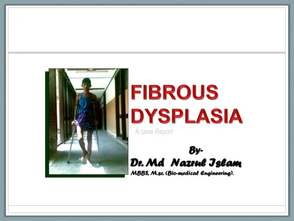
By- Dr. Md Nazrul Islam MBBS, M.sc. (Bio-medical Engineering). FIBROUS DYSPLASIA A case Report
Particulars of the patient • Name: Rabiul Islam • Age: 20 years • Gender: Male • Address: Fulbaria, Bogra • Occupation: Labour • Marital status: Married • Religion: Muslim • Date of admission:17.09.09 • Date of examination:17.09.09
Chief complaints • Pain & deformity at the right upper thigh for 7 months following a trauma. • Gradual shortening of the right lower limb with difficulty in walking for 6 months.
History of present illness According to the statement of the patient, he was reasonably well 7 months back, then suddenly he felt down on the ground by accidental trauma. He could walk following trauma without support, after which he noticed mild, fixed aching pain in the right upper thigh which was not associated with fever, non-radiating & aggravated during walking & incompletely relived by taking some pain killers.
History of present illness…cont He also noticed a deformity in supero-lateral aspect of right thigh which was gradually increasing in size, associated with bending of the affected part & shortening of the lower limb. For which his walking became difficult & was possible only with a support, for the last 6 months.
History of present illness…cont He has neither complain of pain & deformity in the other parts of the body nor H/O weight loss or loss of appetite . With these complaints he got admitted at ShaheedSuhrawardy Medical college Hospital for better management.
History of past illness • He had no history of tuberculosis. • He is non Diabetic
Family history None of his family member suffered from such illness. Personal history He is not smoker
Socio-economic Lower middle class family Immunization history Immunized against tuberculosis & tetanus Drug history H/O taking NSAIDs to relieve pain
General examination • Appearance: Ill looking • Body built: Average • Co-operation: Co-operrative • Decubitus: On choice • Anaemia: Absent • Jaundice: Absent • Cyanosis: Absent • Oedema : Absent • Temperature: normal
General examination…..cont. • Pulse: 76 bts/min • Blood pressure: 110/70 mm of Hg • Respiratory rate: 16 /min • Dehydration: No sign • Koilonychia: Absent • Leukonychia: Absent • Clubbing: Absent • Neck vein: Not engorged • JVP: Not raised • Lymph nodes: Not palpable • Thyroid gland: Not palpable • Skin pigmentation: Absent
Local examination: (Right Upper thigh) Look: • An ill defined deformity occupying at the supero-lateral aspect of the upper right thigh with convexity antero-laterally. • Skin over the deformed area is normal • Varus deformity of hip with shortening of the lower limb. • Unable to walk without support. • Wasting of the thigh, & gluteal muscles • No engorged vein.
Local examination: (Right Upper thigh) Feel: • There is an irregular, expanded bony deformity with convexity antero-laterally extending from the hip to subtrochanteric area. local temperature normal, mild tenderness present, over lying skin is free. • Shortening of limb - 9 cm. • Muscle wasting- Gluteal - 4 cm. Thigh – 4 cm. Leg – 3 cm • Distal neurovascular status normal • Regional lymph nodes not enlarged.
Local examination: (Right Upper thigh) Movement: • walk with support. • Trendelen Burg’s test positive • Right Hip (ROM)– • Flexion 0-1000 [normal 0-1200] • Extension 0-50 [normal 0-200] • Abduction 0-50 [normal 0-400] • Adduction 0-150 [normal 0-250] • Internal rotation at 900 flexion 0-200[0-450] • External rotation at 900 flexion 0-100 [0-450] • Internal rotation in extension – 0-200 [0-350] • External rotation in extension – 0-150 [0-450] • Rt. Knee & ankle: normal range of movement
Systemic examination: Locomotorsystem Gait:Can walk with support Inspection:Varusdeformity - right hip Palpation:Tenderness – affected area Spine:Normal
Nervous system examination • Higher psychic function: Normal • Cranial nerve examination: Normal • Motor function: Inspection: Gross Muscle wasting in right hip, thigh & leg
Palpation: Nervous system examination…cont Bulk of muscle: Wasting Hip-4cm. thigh: 4cm, Leg 3cm Tone of muscle:muscle tone is normal
Nervous system examination…cont. Power: [MRC scale] Hip (rt.): extensor- 2 internal rotator- 4 flexor- 4 external rotator- 3 adductor- 4 abductor- 3 Knee (rt.): extensor- 3 flexor- 3
Nervous system examination…cont. • Deep tendon reflex: All jerks are present & normal • Sensory function test: All the sensory functions are normal
Alimentary system examination • Inspection: nothing abnormality detected • Palpation: soft, non tender • Percussion: tympanic • Auscultation: bowel sound present • Per-rectal examination: normal findings
Respiratory system examination • Inspection: Normal in size & shape of the chest • Respiratory rate: 16 /min • Palpation: Trachea centrally placed, normal chest expansibility • Percussion: Resonant • Auscultation: Bronchial breathing sound with no added sound
Cardiovascular system examination • Pulse: 76 bts/ min • B.P. 110 mm of Hg • JVP: Not raised • Inspection: NAD • Palpation: Apex beat in Lt 5thintercostal space, NAD • Percussion: superficial cardiac dullness present over the precordium • Auscultation: s1 & s2 is audible Geneto - Urinary system examination Reveals no abnormality
Salient feature Mr. Rabiul Islam, a 20 years old man, coming from Fulbaria, Bagura admitted in ShaheedSuhrawardy Medical College Hospital with the complaints of pain & deformity at the rt. Upper thigh following a mild accidental trauma 7 months back & gradual shortening of rt. Lower limb with difficulty in walking for 6 months.
Salient feature….cont. The pain was mild , fixed, non radiating, aching in nature which was not associated with fever, aggravated during walking & incompletely relived by taking NSAIDs. He also noticed a bending deformity in supero-lateral aspect of right thigh which was gradually increasing in size causing shortening of the affected limb
Salient feature….cont. Other parts of the body were normal with no history of weight loss or anorexia. none of his family member suffered from such illness. On general examination, the patient is ill-looking, not anaemic, non icteric, normothermic, normotensive & skin pigmentation is absent.
Salient feature….cont. On local examination, an ill defined, mildly painful bowing deformity was seen occupying at the supero-lateral aspect of the right thigh with convexity antero-laterally extending from the hip to subtrochanteric area with CoxaVara. Overlying skin & local temperature was normal.
Salient feature….cont. Shortening of the limb was found 9 cm than the left. He was unable to walk without support. There was gross muscle wasting in rt. Lower limb, measuring gluteal- 4 cm, thigh- 4cm, leg- 3 cm. with loss of muscle power at hip & knee. Muscle tone was normal.
Salient feature….cont. Distal neurovascular status was normal & Regional lymph nodes were not enlarged. Trendelen Burg’s test was positive with reduced Range of movement (ROM) in hip in all direction. ROM of knee & ankle was normal. The spine was normal. Other systemic examination reveals no abnormality.
Provisional diagnosis ? Fibrous dysplasia – upper third of the right femur
Differential diagnosis • Giant cell tumor • Enchondroma • Aneurysmal Bone Cyst • Brown tumor
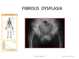
Investigations • X-Ray right thigh with hip A/P & lateral view: Shows Shephard’s crook deformity (neck-shaft angle: 900) with multiple osteolytic lesions involving part of the neck, trochanteric & subtrochanteric area, with thinning of cortical bone & lucent patches typically hazy, looks like ground-glass appearance with pathological fracture at the subtrochanteric region.
Investigations • Blood for • TC of WBC 9,000 / cu mm • DC of WBC • N 56% B 0% • L 26% M 5% • E 4% • ESR 15 mm in 1st hr • Hb% 12 gm / dl • Urine RME Normal study • CXR-P/A view Normal Chest skiagram • MT Not significant • RBS 76 mgm / dl
Investigations • S. creatinine 0.9 mgm/ dl • Blood urea 30 mgm / dl • S. calcium 9 mgm / dl • S. alkaline phosphates 110 IU/ L • FNAC No malignant cell found, only cellular fibrous tissue present.
Confirmatory diagnosis “Monostotic fibrous dysplasia with Shephard’s Crook deformity in upper end of right femur with pathological fracture”
Treatment • This patient was under gone for surgical treatment on 17-10-09 • Procedure: • Through lateral approach upper end of the femur was exposed • Outer part of the proximal femur was so thin that it needs little effort to curate the cystic areas carefully.
Treatment….cont. Procedure…cont.: • After curettage valgus wedge osteotomy was done at subtrochanteric region to correct deformity, massive irradiated allograft with fibular auto graft was applied to enhance healing & incorporation of the cystic bony lesion & fragments were fixed with proximal femoral interlocking nail (PFN).
Treatment….cont. Procedure…cont.: • Wound was closed in layers by keeping a drain inside, which was removed after 48 hrs. • Abduction bar was applied • Specimen was sent for histopathology.
Histop-athologicalReport • Shows loose cellular fibrous tissue with wide spread patches of immature bone - Suggestive of Fibrous dysplasia.
Post operative management& follow up • Stitches were removed after 10th POD • Only isometric quadriceps exercise advised. • He was advised to take calcium& Bisphosphonates preparation regularly. • After removal of the abduction bar at 2 months clinically & radiologically bone was stable & uniting satisfactorily . • Knee bending & quadriceps exercise advised. • He was advised to use crutch for non weight bearing up to 3 months. • After 3 months partial weight bearing started with 2 cm shoe raised along with other exercise.
Last follow up (4 ½ months after surgery) • Clinical • Pain & Deformity markedly reduced • Can walk with single crutch • Muscle power & wasting improving • Now LLD - only 2 cm • Radiological • Deformity is almost corrected • Now neck-shaft angle: 1350 • well incorporation of the grafted bone. • Union process is satisfactory at the osteotomy site.
Peroperative X-ray on 17.10.09 Before & Afterosteotomy
Fig: Post operative X-ray Rt. Upper Femur After 7 weeks On 10th POD
Fig: Post operative X-ray Rt. Upper Femur After 4 ½ months After 3 months