Visual Perception Kit
Visual Perception Kit. Carolina $117.00. Time Required. Approximately 8 minutes per station With 10 stations that will require 80 minutes Additional time is required to answer assessment and review questions. Preparation. Prior to lab review eye structure and function

Visual Perception Kit
E N D
Presentation Transcript
Visual Perception Kit Carolina $117.00
Time Required • Approximately 8 minutes per station • With 10 stations that will require 80 minutes • Additional time is required to answer assessment and review questions
Preparation • Prior to lab review eye structure and function • Lab is designed so that 10 stations can be set up and instructions for each station are included • Pre-lab set-up • Wrap white paper lengthwise around pencils • Set up “Depth Perception Tester” on level surface
Pre-lab Activities • Demonstrate visual acuity test and benefit of corrective lenses • Have students predict and then identify their dominant eye • Look at an object ~3m away • With both eyes open, hold your thumb arms’ length and “cover” the object with your thumb • Close right eye • Results – if your thumb appears to move to the right, the right eye is dominant. If your thumb did not seem to move, your left eyes dominant.
Activities Included • Visual Acuity • Astigmatism and Blind Spot • Visual Mapping • Color Vision • Depth Perception • Accommodation • Near Point • Peripheral Vision • Afterimages • Illusions
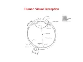
Visual Acuity • Uses Snellen chart (eye chart) to determine emmetropia (normal vision), hyperopia (farsightedness), myopia ( nearsightedness) • Astigmatism and Blind Spot • Astigamatism- irregular shape of cornea or lens • Results in blurred or distorted images • Blind spot- area on retina with no photoreceptors • Visual Mapping • Used to determine the size of the fovea centralis • Fovea centralis- area of eye with greatest visual acuity, contains more cone cells than any other region of eye
Color Vision • Cones- cells in retina which see color • Rods- cells in retina which see dim light and shades of gray • Different cones work together to see different colors • Depth Perception • Ability to judge the relative distance between objects in three dimensions • Done using monocular vision(seeing with one eye) and binocular vision(seeing with two eyes) • Accommodation • Combination of reflex actions by which the lens of the eye changes to keep the focal length constant • If works normally= emmetropia • If eye ball is deeper than normal = myopia • If eye ball is not deep enough= hyperopia
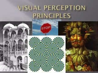
Near Point • Minimum distance at which the eye can focus on an object • Peripheral Vision • Ability to see things which fall outside the direct line of vision • Used in the detection of motion • Afterimages • Optical illusion that occurs when looking away after staring intently at certain images or colors • Illusions • Deceptive, self-contradictory, or misleading images • Visual tricks which take place in the brain rather than the eye
Today… • Get in groups of 3 • We will test our • Depth Perception • Peripheral Vision • Illusions