Download
1 / 34
370 likes | 637 Vues
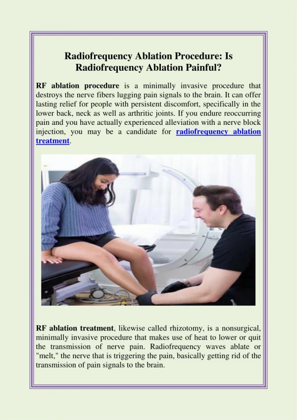
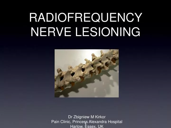
RADIOFREQUENCY NERVE LESIONING. Dr Zbigniew M Kirkor Pain Clinic, Princess Alexandra Hospital Harlow, Essex, UK. 1. Brief history of RF Physics of RF Clinical applications Guidelines and protocols Evidence for efficacy. Brief history of RF Physics of RF Clinical applications

E N D
RADIOFREQUENCY NERVE LESIONING • Dr Zbigniew M Kirkor • Pain Clinic, Princess Alexandra Hospital • Harlow, Essex, UK 1
Brief history of RF • Physics of RF • Clinical applications • Guidelines and protocols • Evidence for efficacy
Brief history of RF • Physics of RF • Clinical applications • Guidelines • Evidence for efficacy
Until 1980 - large size probes (14-gauge) • 1931 - Gasserian ganglion termolesion, • 1975 - RF lesioning of the medial branch for lumbar facets (Shealy), • 1977 - RF lesion of dorsal root ganglion (Uematsu), • cordotomies.
1980 - 1995 - introduction of fine probes • medial branch (facet joints), • dorsal root ganglion, • sympathetic chain, • 1991 - nucleus of the disc.
1996 to present • pulsed RF, • cooled RF, • era of extensive research and search for evidence, • development of computerised generators.
Brief history of RF • Physics of RF / Pulsed RF • Clinical applications • Guidelines • Evidence for efficacy
Your household outlet has AC of 60Hz or 50Hz Radiofrequency Generator - 460kHz = 460,000Hz A microwave - 500-1000kHz Alternating Current and RF AC frequency (f) is the number of cycles per second (measured in Hz)
RF energy is applied Ions in surrounding tissue move creating friction Friction heats surrounding tissue Hot tissue heats probe or electrode by conduction Probe thermocouple located at the tip, reads tissue temperature Ionic Heating Using RF Cannula Exposed Active Tip Insulated Introducer
Temperature drops as radius from tip increases Neurodestruction occurs when temp reaches > 45ºC A small zone of reversible damage surrounds lesion Cannula Insulation Cannula Active Tip 80°C 40°C 65°C Lesion Temperature
Clinical implications • Size matters !!! Larger canniula = larger area of lesion. • Temperature between 45 and 80oC. • Time of lesioning: not less then 60 seconds but not more then 90 seconds. • Tissue impedance: high in dense tissues, low in liquids (blood, CSF, disc). • Probe need to placed parallel (along) to the nerve.
In pulsed RF, the treatment effect is produced by the electromagnetic field. It is not a thermal lesion. Electro-magnetic field is maximal at the tip and decreases behind the tip.
Neuropathic Pain is a contraindication for Standard RF Pulsed RF can treat peripheral nerves without injuring them. Pulsed RF maximizes voltage whilst ensuring that temperature does not exceed 42 For Neuropathic Pain
Brief history of RF • Physics of RF • Clinical applications • Guidelines • Evidence for efficacy
RADIOFREQUENCY • Cervical, thoracic and lumbar facet joints (medial branch), • Sacroiliac joints, • Sympathetic chain.
Pulsed RF • Dorsal root ganglions (nerve roots) - cervical, thoracic, lumbar. • Trigeminal ganglion. • Peripheral nerves.
Brief history of RF • Physics of RF • Clinical applications • Guidelines • Evidence for efficacy
Published in 2004 • Edited by Prof. Nikolai Bogduk on behalf of the Standards Committee of ISIS
ESTABLISHEDPROCEDURES • DIAGNOSTIC • Lumbar spinal nerve blocks • Lumbar disc stimulation • Lumbar medial branch blocks • Cervical disc stimulation • Cervical medial branch blocks
ESTABLISHED PROCEDURES • THERAPEUTIC • Lumbar transforaminal injection of corticosteroids • Percutaneous radiofrequency lumbar medial branch neurotomy • Intradiscal electrothermal therapy • Cervical transforaminal injection of cortycosteroids • Percutaneous radiofrequency cervical medial branch neurotomy
“Lumbar medial branch blocks are diagnostic procedures designed to test if a patient’s pain is mediated by one or more of the medial branches of the lumbar dorsal rami.” • “... Lumbar medial branch blocks are used to test if a patient’s pain stems from a given lumbar zygapophysial (facet) joint. For that purpose the nerves that innervate the joint are anaesthetized.”
Steroid injection into the facet joint is NOT a validated method of treatment. • Diagnostic blocks should be performed twice, ideally as a double blind procedure. • It doesn’t matter what local anaesthetic is used.
Brief history of RF • Physics of RF • Clinical applications • Guidelines • Evidence for efficacy
There is very limited number of well designed Randomized Controlled Trials for radiofrequency neurotomy. • There is no RCT for Pulsed RF neurotomy.
Pain relief was categorized as at least 80% pain relief from baseline pain and ability to perform previously painful movements. For therapeutic interventions, the primary outcome measure was pain relief with secondary outcome measures of improvement in functional status, psychological status, return to work, and reduction in opioid intake. For therapeutic interventions, short-term pain relief was defined as relief lasting 6 months or less and long-term relief as longer than 6 months.
Conclusion: The evidence for diagnosis of lumbar facet joint pain with controlled local anesthetic blocks is Level I or II-1. The indicated level of evidence for therapeutic lumbar facet joint interventions is Level II- 1 or II-2 for lumbar facet joint nerve blocks, Level II-2 or II-3 evidence for radiofrequency neurotomy, and Level III (limited) evidence for intraarticular injections.
Results: Based on the utilization of controlled comparative local anesthetic blocks, the evidence for the diagnosis of cervical facet joint pain is Level I or II-1. The indicated evidence for therapeutic cervical medial branch blocks is Level II-1. The indicated evidence for radio- frequency neurotomy in the cervical spine is Level II-1 or II-2, whereas the evidence is lacking for intraarticular injections.