Sensing the world
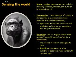
S 1. Sensing the world. Sensory coding : sensory systems code for modality, intensity, location, and duration of external stimuli. Transduction : the conversion of a physical stimulus into a change in membrane potential (electrochemical signal)

Sensing the world
E N D
Presentation Transcript
S 1 Sensing the world • Sensory coding: sensory systems code for modality, intensity, location, and duration of external stimuli. • Transduction: the conversion of a physical stimulus into a change in membrane potential (electrochemical signal) • Signals are transmitted in the form of graded potentials, action potentials, and synaptic interaction • Receptors:cells or regions of cells that respond to specific stimuli and perform transduction • The process of sensory coding starts here • Specificity: receptors are often sensitive to specific stimuli; varies with receptor type
S 2 Changes of membrane potential depend on ion channels Reminder: leak channels are not gated.
S 3 5 different types of receptor proteins (but not all in the same cell) Receptor proteins Receptors for gustation Receptor cell 1st order sensory neuron
S 4 Hair cells = specialized receptor cell with mechanically-gated K+ channels. Receptors for audition (hearing) and equilibrium(movement and orientation to gravity) Receptor cell 1st order sensory neuron Scanning electron micrograph of hair cells from the bullfrog inner ear, which contain the mechanically-gated ion channel TRPA1. (Image courtesy of Howard Hughes Medical Institute)
S 6 Receptor cells withreceptor proteins. Encapsulated TZ TZ TZ
S 7 Somatosensory and the sensation of touch Skin is largest sense organ: up to 2 million receptors Sensory system must code for where (location), how hard (intensity), how long (duration), and modality (sense of touch, temperature, vibration, wet/dry, damage (pain; noxious)).
S 8 Somatosensory: sensation of touch, vibration, pain, and temperature “Adequate” stimulus: the stimulus type to which areceptor responds best (lowest threshold.) Deep Superficial Sustained stimulus Fluctuating stimulus Encapsulated nerve endings
S 9 This diagram is misleading: Different types of receptors are NOT part of the same sensory neuron!
S 10 Labeled Lines: Different sensory modalities are transmitted separately along distinct pathways.
Must Code for 1) Stimulus Localization & 2) Modality & 3) Intensity& 4) Duration S 11 Figure 7.15 Three neurons to the cortex! Labeled Line
S 12 Right side Right side 3 2 Anterolateral (spinothalamic) tract 1st order synapses onto 2nd order in spinal cord, 2nd order axon decussates in spinal cord, travels to thalamus in contralateral anterolateral tract, synpases onto 3rd order neuron in thalamus, which sends its axon to SSC. Dorsal column-medial lemniscal tract 1st order axons ascend in ipsilateral dorsal columns to synapses onto 2nd order in dorsal column nuclei of brainstem, 2nd order axon decussates in brainstem, travel to thalamus to synpases onto 3rd order neuron, which sends it axons to SSC. Right side 1 Right side Proprioception
S 13 Somatotopy in the Somatosensory Cortext (post-central gyrus.) Regions more densely innervated by sensory receptors occupy more cortical tissue.
S 14 Referred pain Who cares? What about referred pain from the heart?
S 16 Referred PainFig 7-18 p. 200 of text
S 17 Receptive field = the area of the body that, when stimulated, leads to activity in a neuron. = 1st order sensory neuron Could be encapsulated
S 18 Right side Right side Receptive field = the area of the bodythat, when stimulated, leads to activity in a neuron. 1st, 2nd, and 3rd order sensory neurons each have receptive fields. Dorsal column-medial lemniscal tract 1st order axons ascend in ipsilateral dorsal columns to synapses onto 2nd order in dorsal column nuclei of brainstem, 2nd order axon decussates in brainstem, travel to thalamus to synpases onto 3rd order neuron, which sends it axons to SSC. Right side Right side Proprioception
S 19 Receptor potentials e.g. somatosensory • Graded potentials are the result of transduction within a receptor. Transduction produces a receptor potential • Amplitude is usually in proportion to the stimulus intensity • Specialty receptor cells with no axon (visual, gustatory, auditory, and vestibular systems). The graded receptor potentials will directly change amount of NT secretion onto 1st order sensory neuron. • Receptors with axons (somatosensory and olfactory systems) have a trigger zone and receptor potentials generate action potentials e.g. gustatory
S 20 Receptors and transduction Could be encapsulated Activation of mechanically gated channels
S 21 • Sensations to touch • Mechanoreceptors contain receptor proteins that respond to stretching, distortion, or pressure on the peripheral plasma membrane
S 23 Summary to now:Modality encoded by _______.Intensity encoded by _______. What’s left?Location encoded by _______.Duration encoded by _______.
Stimulus Localization S 24 Figure 7.06 The size of receptive fields varies dramatically in different regions of skin (i.e. lips, palm, fingertip, calf). For touch discrimination, small receptive fields allow greater accuracy in “two point discrimination” test (in lab!)
Stimulus Localization& Intensity S 25 Receptive fields of different neurons often overlap such that any patch of skin may have several receptors of the same type (modality) and receptors of different types (different modalities I.e. touch, pain temperature, etc.) Overlapping receptive fields of touch receptors (Meissners, Merkels) allow for more precise localization of a stimulus via the mechanism of lateral inhibition (next slide.)
S 26 Stimulus Localization & Intensity Mild inhibition PresynpaticInhibition Massive inhibition Lateral inhibition exaggerates the difference in stimulus intensity detected by adjacent neurons, aids with LOCALIZATION.
Stimulus Localization & Intensity S 27 Lateral inhibition improves stimulus localization. Which is easier to localize and why: a light touch or a moderate touch?
Right side 3 2 Right side 1 Right side Proprioception
1QQ # 12 Answer one • Which of the following are correct statements: a) The intensity of a stimulus is proportional to the size of the graded potential in the receptive membrane.b) The modality of a stimulus is encoded by which type or types of sensory receptors are activated. c) The intensity of a stimulus is encoded by the frequency of action potentials. d) Some ascending spinal neurons receive synaptic inputs from more than one part of the body. e) Each taste receptor cell has at least two different categories of tastant receptor proteins. • What is the meaning of labeled line in the context of sensory physiology?
S 28 Coding for stimulus duration. Adaptation = decrease in frequency of APs with sustained stimulus
S 29 Figure 7.11 “Flutter” receptors are rapidly adapting. Examples: waistband of underwear, top of socks, earrings, mechanoreceptors in carotid arteries for blood pressure YouTube