Download

1 / 45
450 likes | 615 Vues
Skin and Body Membranes. Chapter 4. Body Membranes . Cover surfaces, line body cavities, and form protective (and often lubricating) sheets around organs. Two major groups 1. Epithelial membranes Cutaneous Mucous Serous 2. Connective tissue membranes Synovial. Focus here!.

E N D
Skin and Body Membranes Chapter 4
Body Membranes • Cover surfaces, line body cavities, and form protective (and often lubricating) sheets around organs. • Two major groups • 1. Epithelial membranes • Cutaneous • Mucous • Serous • 2. Connective tissue membranes • Synovial Focus here!
Cutaneous Membrane • Cutaneous membrane = skin • A dry membrane • Outermost protective boundary • Superficial epidermis • Keratinized stratified squamous epithelium • Underlying dermis • Mostly dense connective tissue Figure 4.1a
Mucous Membranes (mucosa) • Surface epithelium • Type depends on site • Underlying loose connective tissue (lamina propria) • Lines all body cavities that open to the exterior body surface (such as respiratory, digestive, urinary, and reproductive tracts) • Often adapted for absorption or secretion • “Wet” or moist membranes Figure 4.1b
Serous Membranes • Surface simple squamous epithelium, Underlying areolar connective tissue • Lines open body cavities that are closed to the exterior of the body • Serous layers separated by serous fluid • Parietal layer: lines a specific portion of the wall of the ventral body cavity • Visceral layer: covers the outside of the organs in the cavity Figure 4.1c
Serous Membranes • Specific serous membranes • Peritoneum • Abdominal cavity • Pleura • Around the lungs • Pericardium • Around the heart Figure 4.1d
Connective Tissue Membrane • Synovial membrane • Connective tissue only (areolar) • Lines fibrous capsules surrounding joints where they provide a smooth surface and secrete a lubricating fluid • Also cushion organs moving against each other during muscle activity, such as the movement of a tendon across a bone’s surface. Figure 4.2
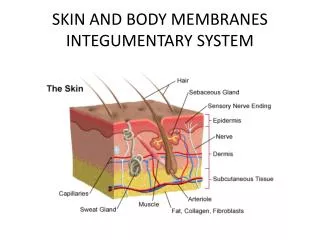
Integumentary System • Skin = cutaneous membrane • The skin and its derivatives serve a number of functions, mostly protective, and together these organs are called the Integumentary System • Skin derivatives • Sweat glands • Oil glands • Hairs • Nails
Skin Functions • Protects deeper tissues from: • Mechanical damage • Chemical damage • Bacterial damage • Thermal damage • Ultraviolet radiation • Desiccation • Aids in heat regulation • Aids in excretion of urea and uric acid • Synthesizes vitamin D Refer to pg.111for the “how?”
Basic Skin Functions Page 111….add three things to your notes that your skin does for you!
Skin Structure • Epidermis – outer layer • Stratified squamous epithelium • Often keratinized (hardened by keratin) • Dermis • Dense connective tissue Figure 4.3
Skin Structure • Deep to dermis is the hypodermis • Not part of the skin • Anchors skin to underlying organs • Composed mostly of adipose tissue • Serves as a shock absorber and insulates the deeper tissues from extreme temperature changes occurring outside the body. • It is also responsible for the curves that are more a part of a woman’s anatomy than a man’s!
Skin Structure Figure 4.4
Layers of EpidermisEpidermis is composed of 5 zones or layers called “strata”…listed below inside out… • Stratum basale • Cells undergoing mitosis • Lies next to dermis • Stratum spinosum • Stratum granulosum • Stratum lucidum • Occurs only in hairless, thick skin like soles of feet and palms of hands • Stratum corneum • Shingle-like dead cells
Epidermis Continued… • Epidermis is avascular…that is has no blood supply of its own • Explains why a man can shave everyday and not bleed even though he is cutting off many cell layers each time he shaves! • Most cells of the epidermis are keratinocytes (keratin cells), which produce keratin, the fibrous protein that makes the epidermis a tough protective layer.
Epidermis Continued…Stratum Basal • Lie closest to the dermis • Contains epidermal cells that receive the most adequate nourishment via diffusion of nutrients from the dermis • Constantly undergoing cell division…millions of cells are produced daily The daughter (new) cells are pushed upward, away from the source of nutrition to become part of the epidermal layers closer to the skin surface (stratum spinosum and granulosum) then become flatter, and finally die, forming the clear stratum lucidum.
Epidermis Continued…Stratum Corneum • The outermost layer • 20-30 cell layers thick! Accounts for three-quarters of the epidermal thickness. • Provides a durable “overcoat” for the body, which protects deeper cells from the hostile external environment (air) and from water loos and helps the body resist biological, chemical, and physical assaults. • Rubs and flakes off slowly and steadily and is replaced by cells produced by the division of the deeper stratum basale cells. • Indeed, we have a totally “new” epidermis every 25-45 days!
The common saying “Beauty is only skin deep” is especially interesting in light of the fact that nearly everything we see when we look at someone is dead!
Dermis • Two layers • Papillary layer (upper dermal region) • Projections called dermal papillae…furnish nutrients to the epidermis • Pain receptors and touch receptors • Capillary loops…distinct looping on hands and feet to help increase friction for gripping ability…also make our unique fingerprints • Reticular layer (deepest skin layer) • Contain blood vessels, sweat and oil glands, and deep pressure receptors
Read pg.114 second column In your notes record Why our skin wrinkles What causes bedsores
Melanin • A pigment that ranges in color from yellow to brown to black • Produced by special cells called melanocytes found chiefly in the stratum basale • When the skin is exposed to sunlight, the melanocytes are stimulated to produce more melanin pigment…then tanning occurs! • The stratum basale cells phagocytize (eat) the pigment as it accumulates in them and the melanin forms a protective pigment umbrella over the superficial side of their nuclei that shields their genetic material from the damaging effects of UV radiation in sunlight. • Freckles and moles are seen where melanin is concentrated in one spot. • But like anything…too much of a good thing….read “homeostatic imbalance” on page 114 and make notes.
Skin Color • Three pigments contribute to skin color: • The amount and kind (yellow, reddish brown, or black) of melanin in the epidermis. • The amount of carotene deposited in the stratum corneum and subcutaneous tissue. Carotene is an orange-yellow pigment found in abundant amounts in carrots and other orange, deep yellow, or leafy green vegetables. The skin tends to take an orange hue when large amounts of carotene-rich foods are eaten. • The amount of oxygen-rich hemoglobin (pigment in red blood cells) in the dermal blood vessels. People who produce a lot of melanin have brown-toned skin. In light skinned (Caucasian) people, who have less melanin, the crimson color of oxygen rich hemoglobin in the dermal blood supply flushes through the transparent cell layers above and gives the skin a rosy glow…when hemoglobin is poorly oxygenated, both the blood and the skin of a Caucasian appears blue (cyanosis) which is common during heart failure and severe breathing disorders.
Skin Color Skin color can also be influenced by emotional stimuli and can also signal certain disease states. • Redness or erythema: • Reddened skin may indicate embarrassment (blushing) fever, hypertension, inflammation, or allergy • Pallor or blanching: • Under certain types of emotional stress (fear, anger, and others), some people become pale. Pale skin may also signify anemia, low blood pressure, or impaired blood flow into the area. • Jaundice: • Yellow cast…an abnormal yellow skin tone usually signifies a liver disorder in which excess bile pigments are absorbed into the blood, circulated throughout the body, and deposited in body tissues (common in newborns) • Bruises: • Black and blue marks reveal sites where blood has escaped from the circulation and has clotted in the tissue spaces. Such clotted blood masses are called hematomas. An unusual tendency to bruising may signify a deficiency of vitamin C in the diet or hemophilia (bleeder’s disease)
Skin Appendages Cutaneous glands, hair and hair follicles, and nails!
Cutaneous Glands • All are exocrine glands that release their secretions to the skin surface via ducts • Fall into two groups: • Sebaceous (oil) Glands • Sweat Glands
Sebaceous (Oil) Glands • Found all over the body, except on the palms of the hands and the soles of the feet. • Their ducts usually empty into a hair follicle, but some open directly onto the skin surface • Product of sebaceous glands: • Sebum…a mixture of oily substances and fragmented cells • Sebum is a lubricant that keeps the skin soft and moist and prevents the hair from becoming brittle • Sebum also contains chemicals that kill bacteria, so it is important in preventing the bacteria on the skin surface from invading the deeper skin regions. • The sebaceous glands become very active when male sex hormones are produced in increased amounts (in both sexes) during adolescence…leading to oilier skin during this period of life.
Sebaceous (Oil) Glands • Read “homeostatic imbalance” on page 117 and answer: • What is a whitehead? • What is a blackhead? • What is seborrhea and why does it occur?
Sweat Glands • AKA: Sudoriferous Glands • Widely distributed in the skin • Their number is staggering-more than 2.5 million per person • Two types: • Eccrine • Far more numerous, Open via duct to pore on skin surface, produce sweat • Apocrine • Ducts empty into hair follicle, begin functioning during puberty, precise function is not yet known, but are activated by nerve fibers during pain and stress and during sexual foreplay.
Sweat and Its Function • Composition • Mostly water plus some salt • Some metabolic waste (ammonia, urea, uric acid) • Is Acidic (pH range 4-6)… a characteristic that inhibits the growth of bacteria! • Function • Helps dissipate excess heat…when sweat evaporates off the skin’s surface, it carries large amounts of body heat with it • Excretes waste products
Hair There are millions of hairs scattered all over the body, but other than serving a few protective functions (head on hair protects head and eyelashes protect eyes), hair has lost much of its usefulness! It once kept early human’s bodies insulated, but now we have other means of keeping warm.
Hair • Produced by a hair follicle, is a flexible epithelial structure • The part of the hair enclosed in the follicle is called the root. • The part projecting from the surface of the scalp or skin is called the shaft. • A hair is formed by division of the well-nourished stratum basale epithelial cells in the matrix (aka: growth zone), of the hair bulb at the inferior end of the follicle. • As the daughter cells are pushed farther away from the growing region, they become keratinized and die. Thus the bulk of the hair shaft is dead material and almost entirely protein.
Hair Anatomy • Central medulla • Cortex surrounds medulla • Cuticle on outside of cortex • Most heavily keratinized • The cuticle is formed by a single layer of cells that overlap one another like shingles on a roof…this design keeps hairs apart and keeps it from matting. • Because the cuticle is most subject to abrasion, it tends to wear away at the tip of the shaft, allowing the keratin fibrils in the inner hair regions to frizz out (aka: split ends!) Hair pigment is made by melanocytes in the hair bulb, and varying amounts of different types of melanin combine to produce all varieties of hair color from pale blond to pitch black.
Hair Fact: • Small bands of smooth muscle cells-arrectorpili-connect each side of the hair follicle to the dermal tissue. • When these muscles contract (like when we are cold or frightened), the hair is pulled upright, dimpling the skin surface with “goose bumps”. • This action helps keep animals warm in winter by adding a layer of insulating air to the fur! • It is especially dramatic in a scared cat, whose fur actually stands on end to make it look larger to scare off its enemy! • However, this hair-raising phenomenon is not very useful to human beings.
Nails • Scale-like modifications of the epidermis that corresponds to the hoof or claw of other animals. • Heavily keratinized • Stratum basale extends beneath the nail bed • Responsible for growth • Lack of pigment makes them colorless (look somewhat pink due to the rich blood supply in the underlying dermis).
Nail Structures Free edge: what hangs past skin Body: visible attached portion Lunula: (lunul=crescent) the half moon near the cuticle. Proximal Nail Fold: Aka-cuticle, projects onto the nail body Nail matrix: Responsible for nail growth Figure 4.9
Developmental Aspects of Skin and Body MembranesFetal Development & Newborns • During the 5th and 6th months of fetal development-the soon to be born infant is covered with a downy type of hair called Lanugo, but this hairy cloak has usually been shed by birth. • When a baby is born, its skin is covered with VernixCaseosa. This white, cheesy-looking substance protects the baby’s skin while it is floating in its water-filled sac inside the mother. • Newborn’s skin is very thin, and blood vessels can easily be seen through it. • Commonly, there are accumulations in the sebaceous glands, which appear as small white spots called Milia on the baby’s nose and forehead…these usually disappear by the third week after birth.
Developmental Aspects of Skin and Body MembranesAdolescence & Young Adults • The skin and hair become more oily as sebaceous glands are activated, and acne may appear…..which usually subsides in early adulthood. • Skin reaches its optimal appearance when we are in our twenties and thirties. • Then visible changes in the skin begin to appear as it is continually assaulted by abrasion, chemicals, wind, sun, and other irritants and as its pores become clogged with air pollutants and bacteria. • As a result, pimples, scales, and various kinds of dermatitis, or skin inflammation, become more common.
Developmental Aspects of Skin and Body MembranesOld Age • The amount of sebaceous tissue decreases, leading to the intolerance to cold so common in the elderly. • The skin also becomes drier, and as a result it may become itchy and bothersome. • Thinning of the skin, another result of the aging process, makes it more susceptible to bruising and other types of injuries. • The decreasing elasticity of the skin, along with the loss of subcutaneous fat, allows bags to form under our eyes, and our jowls begin to sag…this loss of elasticity is sped up by smoking and sunlight. • Although there is no way to avoid the aging of the skin, good nutrition, plenty of fluids, and cleanliness help delay the process.
Developmental Aspects of Skin and Body MembranesHair Loss, Baldness, and Graying… • Page 124 • Second Column • Second Paragraph Add to your notes why we bald and go gray. And, can we regain hair in certain scenarios?
Homeostatic Imbalances of Skin: • The skin can develop more than 1000 different ailments. • The most common skin disorders result from allergies or bacterial, viral, or fungal infections. • Less common, but far more damaging, are burns and skin cancers. • Some Examples: athlete’s foot, boils, cold sores, impetigo, psoriasis, etc…
Skin Homeostatic Imbalances • Burns • Tissue damage and cell death caused by heat, electricity, UV radiation, or chemicals • Associated dangers • Dehydration • Electrolyte imbalance • Circulatory shock
Rule of Nines • Way to determine the extent of burns • Body is divided into 11 areas for quick estimation • Each area represents about 9% Figure 4.11a
Severity of Burns • First-degree burns • Only epidermis is damaged • Skin is red and swollen • Sunburn is an example • Second degree burns • Epidermis and upper dermis are damaged • Skin is red with blisters • Third-degree burns • Destroys entire skin layer • Burn is gray-white or black
Critical Burns • Burns are considered critical if: • Over 25% of body has second degree burns • Over 10% of the body has third degree burns • There are third degree burns of the face, hands, or feet