Download
1 / 57
710 likes | 1.42k Vues
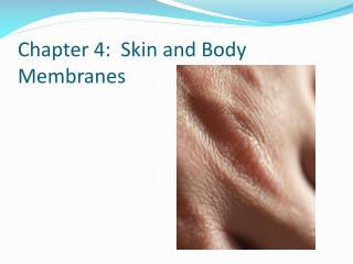
Chapter 4: Skin and Body Membranes. Skin and Body Membranes. Remember an organ is where 2 or more kinds of tissues work together to perform some specific function SKIN: epithelial, connective, muscle and nervous tissue Integument: the membranes of the body that cover, line and protect.

E N D
Skin and Body Membranes • Remember an organ is where 2 or more kinds of tissues work together to perform some specific function • SKIN: epithelial, connective, muscle and nervous tissue • Integument: the membranes of the body that cover, line and protect. • Includes skin, hair, nails and other membranes
Types of Body Membranes • Epithelial membranes • Cutaneous membranes • Mucous membranes • Serous membranes • Connective tissue membranes • Synovial membranes
Serous Membranes • Lines body cavities—covers organs in the thorax and abdomen • Consists of simple squamous epithelial tissue and a thin layer of connective tissue • 2 layers of epithelium • Visceral layer covers the outside of the organ • Parietal layer lines a portion of the wall of ventral body cavity
Serous Membranes Cells of this membrane secrete a clear fluid (serous) between the two layers which helps to lubricate the organs and cavities they are in
Serous Membranes Specific serous membranes Peritoneum -Abdominal cavity Pleura -Around the lungs Pericardium -Around the heart
Mucous Membranes Line body cavities that are open to exterior Ex: nasal and oral cavities Also in tubes of the respiratory, digestive, urinary and reproductive systems. Cells in this membrane secrete mucus Goblet cells make mucus
Cutaneous Membrane Cutaneous membrane = skin
Connective Tissue Membrane Synovial membrane Found in the linings of the joint cavities between the ends of bones Secretes a thick colorless fluid that lubricates the joint
Integumentary System • Functions: Protective covering, regulates body temperature, contains sense organs and excretes waste. • Produces Vitamin D in response to UV radiation. This helps the body absorb calcium
Skin Layers Epidermis—outer layer Stratified squamous epithelium Sheds every 30 days Dermis-inner layer Thicker than epidermis Includes connective and nervous tissue as well as muscle tissue
Skin Layers • Subcutaneous (hypodermis) • Not really considered a layer of skin but found underneath the dermis is deep to dermis • Anchors skin to underlying organs • Composed mostly of adipose tissue • Blood vessels for skin are here
Epidermis • Lacks blood vessels • Cells at very top are far away from blood vessels below and eventually die. • They still provide protection even though they are not functional cells • These cells have keratinized • Keratin is a waterproof protein that forms in the skin cells as they mature
Epidermis • The epidermis shields the underlying tissue from water loss and injury • Also protects against invasion of microbes and bacteria • Melanin is found here. • Melanin is a dark pigment produced by melanocytes • The job of melanin is to absorb light energy to protect skin from damage.
Epidermis • If your skin does not have enough melanin you are at risk for a sunburn. • We all have the same number of melanocytes but genetics determine how much melanin our cells produce
Epidermis • Those people with dark skin have ancestors that live near the equator or at the poles. (both places have periods of direct sunlight) • Color is yellow to brown to black
Epidermis Skin color is also determined by oxygen content. Most of us have a pinkish cast to our skin because of high oxygen Person with oxygen poor blood because of disease or injury have a bluish cast to their skin. This is called cyanosis
Stratum Basale; lowest layer of epidermis • Mitosis takes place here • Stratum corneum; top layer of epidermis • All cells here are dead
Dermis • Binds epidermis to the subcutaneous layer • Made of fibrous connective tissue, blood vessels, nerve fibers, muscle tissue, sebaceous (oil) glands and sweat glands • Thickest layer of skin • Collagen and elastin: fibers that give skin elasticity (ability to return to original shape) and extensibility (ability to stretch) • Lack of collagen and elastin = wrinkles! • Papillary layer (all dermal papillae) causes fingerprints
SPF-Preventing sun damaged skin What is SPF? Is a higher SPF better? Are tanning beds safe?
Accessory Organs--Hair Consists of hard keratinized epithelial cells Hair follicle extends from dermis to surface of skin. Contains the root Hair growth—scalp hair grows for about 3 years, then rests for one Normal hair loss per day is about 100 hairs Androgens are necessary for hair growth, but too much can cause hair loss (male pattern baldness)
Accessory Organs--Hair Melanocytes provide pigment for hair color Hair color is determined by the amount of melanin produced by the melanocytes arrector pili muscle: attached to the hair follicle. When it contracts it causes the hair to stand on end. Goose bumps!
Accessory Organs—Sebaceous Glands • Sebaceous glands: associated with hair follicle, produce oily secretion called sebum. • Sebum: mixture of fatty material and dead cells—keeps hair and skin soft to prevent dry and cracked skin. • Hair follicle with sebum stuck in them= blackhead • Black is oxidized oil—not dirt • If bacteria present can form pimple or boil
Accessory Organs—Sweat Glands • Sweat glands: sudoriferous glands that occur in the skin • They originate in the dermis
Accessory Organs—Sweat Glands • Two types of sweat glands: • Eccrine: associated with pores, produce sweat in response to increased body temperature: begin working right after birth, found all over the body • Apocrine: associated with hair follicles produce sweat in response to stress; begin working at puberty. Contains a higher fat content than normal sweat. Found mainly in armpits, genital area and around breasts. Theorized that this sweat contains pheromones. (thicker and stickier than regular sweat)
Sweat and Its Function • Composition • Mostly water • Salts and vitamin C • Some metabolic waste • Fatty acids and proteins (apocrine only) • Function • Helps dissipate excess heat • Excretes waste products • Odor is from associated bacteria
Accessory Organs—Nails • Scale-like modifications of the epidermis • Heavily keratinized • Lack of pigment makes them colorless • Body is the visible attached portion • Root of nail embedded in skin • Cuticle is the proximal nail fold that projects onto the nail body
Clinical Application • The pink color of the nail bed is clinically significant in that it can aid in the assessment of perfusion (blood flow) to the extremities and can be a determinant of oxygenation. If you pinch one of your fingernails straight down with the thumb and index finger for 5 seconds you will note that your nail bed went from a blanched white color back to pink in a matter of seconds (good profusion). If it takes longer than 3 seconds for the nail bed to “pink up” then profusion to the extremities is considered sluggish. This test is called capillary refill time.
Regulation of body temperature • As body temp increases blood vessels dilate and carry hot blood to the surface to release heat to the atmosphere. • Sweat glands (eccrine) send sweat to the surface to cool the body through evaporation.
Regulation of body temperature • As body temperature decreases blood vessels constrict to prevent blood from flowing to the surface, thereby conserving heat • Shivering also occurs; muscle contractions produce heat through friction • Arrectorpili muscles contract.
3 Input: Information sent along afferent pathway to Brain The Integumentary System: Negative Feedback 4 Output: Information sent along efferent pathway to activate Blood Vessels Sweat Glands Hypothalamus Change detected by receptor 2 Blood vessels dilate causing heat to radiate from the body Sweat Glands become active, cooling through evaporation. 5 1 Stimulus: Increased body temperature Imbalance Body Temperature Imbalance
3 Input: Information sent along afferent pathway to Brain The Integumentary System: Negative Feedback 4 Output: Information sent along efferent pathway to activate Blood Vessels Sweat Glands Muscles Hypothalamus Change detected by receptor 2 Blood vessels constrict Sweat Glands become less active, muscles Shiver to generate heat 5 1 Stimulus: Decreased body temperature Imbalance Body Temperature Imbalance
Skin Homeostatic Imbalances • Macule-discolored spot; freckle • Wheal-localized elevation of the skin that is often accompanied by itching; contact dermatitis
Skin Homeostatic Imbalances • Papule-solid circumscribed, elevated area on the skin; pimple • Nodule- larger papule; acne vulgaris
Skin Homeostatic Imbalances • Vesicle-small fluid filled sac; blister (a bulla is a larger one; chicken pox) • Comedo-yellow or black plug in the skin; blackhead
Skin Homeostatic Imbalances • Pustule-small, elevated, circumscribed lesion of the skin that is filled with pus; whitehead • Erosion (Ulcer)-an eating or knowing away of tissue; decubitus ulcer
Skin Homeostatic Imbalances • Crust-dry, serous, brown, yellow, red or green exudation; eczema • Scale-thin, dry flake of epithelial cells; psoriasis
Skin Homeostatic Imbalances • Fissure-crack like sore or slit that extends through the epidermis into the dermis; athlete’s foot • Cyst-closed sac under the skin; sebaceous cyst
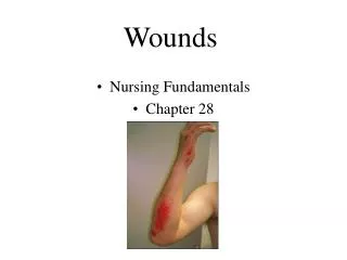
Skin Homeostatic Imbalances • Burns • Tissue damage and cell death caused by heat, electricity, UV radiation, or chemicals • Associated dangers • Dehydration • Electrolyte imbalance • Circulatory shock
Rule of Nines • Way to determine the extent of burns • Body is divided into 11 areas for quick estimation • Each area represents about 9% of total body surface area
Severity of Burns • First-degree burns • Only epidermis is damaged • Skin is red and swollen • Second-degree burns • Epidermis and upper dermis are damaged • Skin is red with blisters • Third-degree burns • Destroys entire skin layer • Burn is gray-white or black
Severity of Burns Figure 4.11b
Critical Burns • Burns are considered critical if • Over 25% of body has second-degree burns • Over 10% of the body has third-degree burns • There are third-degree burns of the face, hands, or feet
Skin Cancer • Cancer—abnormal cell mass • Classified two ways • Benign • Does not spread (encapsulated) • Malignant • Metastasized (moves) to other parts of the body • Skin cancer is the most common type of cancer