Download
1 / 42
420 likes | 698 Vues
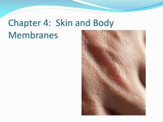
Skin and Body Membranes. Classification of Body Membranes. Body membranes cover surfaces, line body cavities, and form protective sheets around organs The two major groups are epithelial and connective tissue. Classification of Body Membranes.

E N D
Classification of Body Membranes • Body membranes cover surfaces, line body cavities, and form protective sheets around organs • The two major groups are epithelialand connective tissue
Classification of Body Membranes • Epithelial membranes include the cutaneous (skin), mucous, and serous membranes • These membranes are actually simple organs, because they contain an epithelial layer and an underlying connective layer
Classification of Body Membranes • Cutaneous Membrane • The skin • Unlike other epithelial membranes, the cutaneous membrane is a dry membrane
Classification of Body Membranes • Mucous Membranes (mucosa) • Composed of epithelium resting on a loose connective membrane tissue (lamina propria) • Lines all body cavities that open to the exterior • Moist membranes • The epithelium is often adapted for absorption or secretion – not necessarily of mucous!
Classification of Body Membranes • Serous Membranes (serosa) • Composed of simple squamous epithelium resting on a thin layer of areolar tissue • Line body cavities closed to the exterior • Occur in pairs – parietal layerlines specific portion of the wall of the vental body cavity. It folds in to form the visceral layer, which covers the outside of the organs in that cavity
Classification of Body Membranes • Separated by serous fluid • Specific names: • Peritoneum (abdominal cavity) • Pleura (lungs) • Pericardium(heart)
Classification of Body Membranes • Synovial Membranes • Composed of connective tissue – no epithelial cells • Line the capsules surrounding joints and connective tissue that cushion organs
Integumentary System • Integumentary System • Composed of the skin, sweat and oil glands, hairs, and nails • Serves a number of functions, mostly protective
Basic Skin Functions • Integument (Skin)Functions • Keeps water and other essential molecules in the body • Insulates and protects deeper body organs • Protects entire body from mechanical, chemical, and thermal damage, uv radiation, and bacteria • Regulates heat loss • Excretes waste
Basic Skin Functions • Manufactures immunity proteins • Synthesizes vitamin D • Touch, pressure, temperature, and pain receptors provide information about external environment
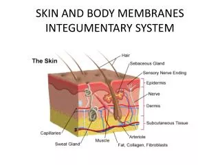
Structure of the Skin • Epidermis • Made up of stratified squamous epithelium that is capable of keratinizing (hardening)
Structure of the Skin • Dermis • Made of dense connective tissue • Below the epidermis and firmly connected to it • Burns or friction cause the dermis and epidermis to separate, leading to blisters
Structure of the Skin • Subcutaneous Tissue (hypodermis) • Adipose (fat) tissue • Not considered part of the skin, but anchors the skin to underlying organs • Serves as a shock absorber • Insulates the deeper tissues from extreme temperatures
Structure of the Skin • Epidermis • Made of five zones/layers called strata • Like all epithelial tissues, the epidermis is avascular (no blood supply of its own) • Most cells are keratinocytes that produce keratin
Structure of the Skin • Stratum Basale (stratum germinativum) • Lies closest to the dermis • Contains epidermal cells that receive nourishment via diffusion from the dermis • Cells are constantly undergoing cell division – millions are produced daily • Daughter cells are pushed upward to become part of the epidermal layers closer to the skin surface
Structure of the Skin • Stratum Spinosum & Stratum Granulosum • More superficial layers of the epidermis
Structure of the Skin • Stratum Lucidum • Cells become flatter and increasingly full of keratin and finally die to form this layer • This layer not seen in all skin regions – occurs only where the skin is hairless and extra thick (palms of hands and soles of feet) • The more superficial layers of the epidermis are “doomed” because they are unable to get inadequate nutrients and oxygen
Structure of the Skin • Stratum Corneum • The outermost epidermis layer – 20 to 30 cell layers thick; accounts for about 75% of epidermal thickness • Made of dead cell remnants, completely filled with keratin (cornified or horny cells) • Protects deeper cells from external environment and water loss • Flakes off slowly and steadily and is replaced by the cells produced in the stratum basale– have a totally “new” epidermis every 25-45 days
Structure of the Skin • Melanocytes • Found chiefly in stratum basale • Produce melanin, a brown-yellow pigment • When exposed to sunlight, more melanin is produced. The cells in the stratum basale “eat” the pigment and as it accumulates in those cells it forms a protective shield for the DNA in the skin cells • A concentration of melanin = moles, freckles
Structure of the Skin • Dermis • Strong, stretchy envelope that helps hold the body together • Consists of two major regions, papillary and reticular • Varies in thickness (eg, thick on palms, thin on eyelids)
Structure of the Skin • Papillary Layer • Upper dermal region • Uneven and has fingerlike extensions from its surface (dermal papillae) which indent the epidermis above • Many contain capillary loops that provide nutrients to the epidermis • Some have pain receptors (free nerve endings) or touch receptors (Meissner’s corpuscles)
Structure of the Skin • Reticular Layer • Deepest skin layer • Contains blood vessels, sweat & oil glands, and deep pressure receptors (Pacinian corpuscles) • Phagocytes act to prevent bacteria from penetrating deeper into the body
Structure of the Skin • Collagen & elastic fibers found throughout the dermis • Dermis is abundantly supplied with blood vessels that play a role in maintaining body temperature homeostasis • Has a rich nerve supply
Skin Color • Three pigments contribute to skin color: • Melanin – increased amount means darker skin (yellow, reddish brown, black) • Carotene – in stratum corneum & subcutaneous tissue (orange-yellow) • Hemoglobin – increased amount in dermal blood vessels increases redness (pigment in red blood cells)
Skin Color • Skin color is influenced by emotional stimuli and color alterations could signal certain disease states • Erythema(redness) – reddened skin may indicate embarrassment, fever, hypertension, inflammation, allergy • Pallor/Blanching – may be due to certain types of emotional stress, anemia, low blood pressure, impaired blood flow
Skin Color • Jaundice (yellowing) – usually signifies liver disorder in which excess bile pigments are absorbed in the blood and circulated throughout the body • Bruises (black and blue) – sites where blood has clotted in tissue spaces (hematomas). An unusual tendency to bruise may be due to vitamin C deficiency or hemophilia
Appendages of the Skin • The skin appendagesinclude cutaneous glands, hair, hair follicles, and nails • Cutaneous Glands • All exocrine glands • Formed by the cells of the stratum basale, and pushed into the dermis • Two types – sebaceous and sweat
Appendages of the Skin • Sebaceous (Oil) Glands • Found all over the skin (except palms and soles) • Their ducts usually empty into a hair follice • Their product is sebum, a mixture of oil substances and fragmented cells • Keeps skin soft and moist and keeps hair from becoming brittle • Contains chemicals that kill bacteria • Become very active when male sex hormone production is increased
Appendages of the Skin • Sweat (sudoriferous) Glands • Widely distributed in skin (about 2.5 million per person) • Two types – eccrine and apocrine
Appendages of the Skin • Eccrine Glands • Found all over the body • Produce sweat, a clear secretion that is primarily water plus salts, vitamin C, trace metabolic waste, and lactic acid • Sweat is acidic (pH 4-6) which inhibits bacteria growth • Reaches skin surface through a pore • Have nerve endings that cause them to secrete sweat when body temperature or external temperature is high
Appendages of the Skin • Apocrine Glands • Largely confined to the axillary and genital areas • Larger than eccrine glands and ducts empty into hair follicles • Secretions contain fatty acids and proteins in addition to substances found in sweat – odorless, milky/yellowish color • Begin to function during puberty under the influence of androgens • Precise function not yet known
Appendages of the Skin • Hairs • Millions scattered all over the body • Few minor protective functions but has mostly lost its usefulness in humans • Produced by a hair follicle (flexible epithelial structure) • The part of the hair enclosed in the follicle is the root • The part projecting from the surface of the skin is the shaft
Appendages of the Skin • Formed by division of stratum basale epithelial cells in the matrix (growth zone)of the hair bulb at the inferior end of follicle • As daughter cells are pushed away from the matrix, they become keratinized • The bulk of the hair shaft is dead and mostly protein
Appendages of the Skin • Each hair consists of a central core called the medulla surrounded by a bulky cortex layer. • The cortex is enclosed by a cuticle formed by a single layer of cells that overlap like shingles. It is the most heavily kertainized region and provides strength and keeps the inner hair layers tightly compacted • Tends to wear away at the tip of the shaft = split ends • Hair pigment is made in the hair bulb
Appendages of the Skin • Hairs come in a variety of shapes and sizes • Humans are born with as many hair follicles as they will ever have • Hairs are among the fastest growing tissues in the body • Hormones account for the development of hair regions
Appendages of the Skin • Hair folliclesare compound structures • The epidermal sheath is composed of epithelial tissues and forms the hair • The outer dermal sheath is dermal connective tissue that supplies blood vessels to the epidermal portion and the papilla provide blood supply to the matrix of the hair bulb • Small bands of smooth muscle cells (arrectorpili) connect each side of the hair follicle to dermal tissue
Appendages of the Skin • Nails are scalelike modifications of the epidermis • Each nail has a free edge, a body (visible attached portion) and a root (embedded in the skin) • The borders are overlapped by skin folds (nail folds). The thick proximal nail fold is called the cuticle
Appendages of the Skin • The stratum basale of the epidermis extends beneath the nail (nail bed) • Its thickened proximal area (nail matrix) is responsible for nail growth • As nail cells are produced by the matrix, they become heavily keratinized and die • Nails are transparent but look pink because of rich blood supply in underlying dermis. The exception is the area over the nail matrix (lunula) that is a white crescent
Developmental Aspects of Skin • During the 5th and 6th months of fetal development the fetus is covered in a downy type of hair (lanugo) but this is shed by birth • At birth, babies are covered in vernixcaseosa (white substance) that protects the babies skin while in the amniotic sac • Newborn skin is very thin and blood vessels can be seen through it. As the baby grows, skin becomes thicker and moist and more subcutaneous fat is deposited
Developmental Aspects of Skin • During adolescence, skin and hair become more oily • Skin’s optimal appearance is during the 20s • During old age, the amount of subcutaneous tissue decreases and skin become drier and more susceptible to bruising • Elasiticity decreases causing sagging and wrinkles
Developmental Aspects of Skin • Hair loses luster with age and by age 50 the number of hair follicles have decreased by 1/3 and continues to decline • Graying hair is caused by a “delayed action” gene. Once the gene takes effect, the amount of melanin deposited in hair decreases or becomes absent