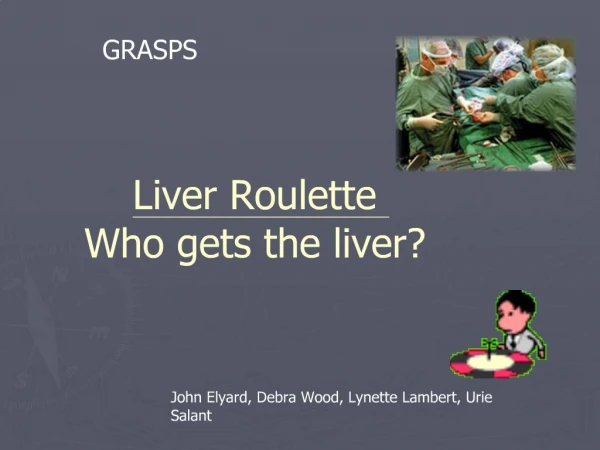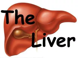
The liver
The liver. Dr. Mezjda Ismail Rashaan,consultant surgeon University of Sulaymania Faculty of medical sciences School of medicine Kurdistan. Anatomy and embryology of the liver:-. -foregut structure, endodermal bud(liver, gal bladder, extrahepatic ducts

The liver
E N D
Presentation Transcript
The liver Dr. Mezjda Ismail Rashaan,consultant surgeon University of Sulaymania Faculty of medical sciences School of medicine Kurdistan
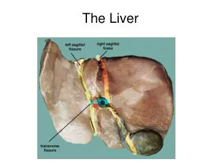
Anatomy and embryology of the liver:- -foregut structure, endodermalbud(liver, gal bladder, extrahepatic ducts -liver cells are bipotential develop(hepatocytes & intrahepatic bile ducts cells) -liver endothelial cells arise from vitellian & umbilical vein, this form sinusides -Glissons capsule -ligaments include: .rt & lt triangular lig. .falciformlig. (from umbilicus to interlober fissure of liver) .coronary lig. .hepatoduodenallig. .gastrohepaticlig.
Blood supply and neural innervations:- -blood supply: .75% portal vein .25% hepatic artery from: 80% caeliac art. 20% SMA .venous (rt., lt, middle hepatic vein to IVC) -neural is via parasympathetic (vagus nerve)and sympathetic
Lymphatics drainage:- -peri-sinusidalspace of Disse and peri-portal clefts of Mall to hilar cystic duct LNs and with common bile duct to caeliac area…
Internal structure:- -8 segmental functional units: .right include 5,6,7,8 segments .left include 1,2,3,4 segments -real functional unites are lobules - Cantlies line separate rt & lt lobes
Functions of the liver:- (storage, metabolism, production, secretion) 1-maintain core body temp. -Ph balance and correction of the lactic acidosis 3-synthesis of clotting factors 4-glucose metabolism, glycolysis, glycogenesis 5-protein metabolism, urea formation 6-bilirubin formation 7-drugs and hormones metabolism 8-removal of gut endo-toxins & foreign bodies.. (reticuloendothelial system)
Investigations:- 1-LFT .bilirubin ( pre-, post- & hepatic dis.) .alk. phosphatase(obstructive jaundice, cholestatic liver) .AST,ALT ( increase in acute hepatocellular dis. like viral hepatitis, alcoholic abuse, autoimmune dis., medications…) .GGT (liver injury , acute alcohol ingestion) .Albumin .prothrombine time ( PT )
2- Imaging:- A-Sonography .liver tumor, bile duct dilatation, gal stones .dopplersonography flow of HA, PV, HV .guiding percutaneouse biopsy .therapeutic as in abscess drainage by pig- tail catheter
Invest. Cont. B- CT –Abdomen with or without contrast ( oral & intravenouse contrast) .lesions .haemangiomas .inflammatory ring enhancement .density (solid or cystic lesions)
Invest.cont:- C-MRI .no iodine .non invasive D-MRCP E-MRA .for chronic liver disease and coagulopathy PV thrombosis
Ivest.cont:- E- ERCP .in obstructive jaundice .stone retrieval .balloon dilatation of stricture .endoprothese .brush cytology F-EUS .hilar tumor extend
Invest. Cont:- G-PTC .if ERCP failed or impossible as in patient with previous polyagastrectomy, in hilar bile duct tumor
Invest. Cont:- H-Angiography .selective for diagnosis and therapeutic .visualize rt, Lt. hepatic art. .patency of portal vein .nature of liver nodule as primary liver tumor has good arterial blood supply .therapeutic intervention as in :- -embolisationof bleeding sites -occlusion A-V malformation -treatment of liver tumor ( chemoembolisation)
Invest. Cont:- I-Nuclear medicine scanning with Iodoida is Te 99 labelledredionuclidespecially in diagnosis of bile duct leak, biliary obstruction. -Sulpher colloid liver screening for kupffer cells, in adenoma and haemangioma no kupffer cells so it is not enhanced
Invest. Cont:- J-Laparascopic & Laparascopic US .staging of hepatopancreaticobiliary cancer which not seen by other methods . In 30% to diagnose peritoneal metas. and superficial liver tumors . With US increase this % by showing also the relation of the tumor to the bile ducts art.
Invest.cont:- K-Flurodeoxyglucose-positron emission tomography ( FDH-PET) . Depend on glucose intake by cancer cells in comparison with adenoma, liver inflammation.
Liver trauma:- -may be blunt or penetration type. -diagnosis depend on clinical suspicion. 1-all lower chest and upper abdominal stab 2-sever crush injuries of no 1 + # ribs, haemo-, pneumothorax 3- penetrating wounds 4-patient with blunt trauma an haemo-dynamically stable but has objective sings as upper abdominal tenderness& gardening 5-peritoneal lavage bloody 6-by laparascopy
Initial management of liver injury:- 1- Penetrating .resuscitation, ABC principles of ATLS. .two large bore cannula .cross matching of blood ( FFP, cryoprecipitate ) .full blood count .LFT, electrolytes, urea , glucose, amylase, clotting screen measurement .arterial gas analysis .chest tube if indicated .transfer the patient to theater
2-Blunt trauma:- -same as penetrating wounds -if stable do imaging for nature of the injury -some cases can be treated conservatively but penetrating needs always operation -indication to do operation in blunt trauma 1-ongoing bleeding 2-coagulopathy 3-generalized peritonitis
Surgical approach:- -rooftop incision -Pringle maneuver -AB -treatment further depend on the type of the injury -damage control surgery by packing in sever injuries
Complications of liver injuries:- 1-sudden massive blood lose 2-delay He 3-subcapsular & intrahepatichaematoma 4-liver abscess duo to liver ischemia or seroma & haematoma infection 5-biliary fistula causing peritonitis 6-haemobilia causing upper rt. Quaderant pain, upper GIT bleeding, jaundice 7-hepatic artery aneurysm 8-arteriovenouse fistula 9-arteriobiliary fistula 10-portovenouse hypertention if aneurysm ruptured to portal vein 11-biliovenouse fistula causing jaundice 12-bronchiobiliary or pleurobiliary fistula 13-liver failure in extensive liver trauma
Long term outcome of liver trauma 1-liver parenchyma regeneration occur 2-biliary tract stricture may be . segmental or lobular needs conservative treatment .or dominant extra-hepatic bile duct stricture causing obstructive jaundice treated by endo-biliary ballooning or stenting or Roux-en-Y hepatodochojejunostomy
Liver cysts May be :- 1-primary congenital .5-14% .as simple cysts or polycystic liver disease .common in females 2-secondary duo to :- .trauma .infections( pyogenic or paracytic) .neoplastic
1-simple cystic lesions -common incidental sonographic finding -asymptomatic -needs no treatment -large one if causing abdominal discomfort do aspiration, if reoccur do deroofinglaparascopic or open laparatomy
2-polycystic liver disease -congenital one associated with other organs as pancrease,kidneys -asymptomatic and incidentalysonographic finding -no effect , no treatment -If multiple cyst causing discomfort give simple pain killer ,if not responding or causing sever pain which is duom to He to the cyst do laparascopic or open fenestration of the cyst.
3-Hydatid liver disease -common in Mediterranean countries -common in liver (70%), long,brain, bones …. -Echinococcusgranulosum -Humanbeing is its interm. Host. ingestion of ova pass to intestine ,portal vein ,liver (larval or cystic stage) -
Clinical features:- -silent seen by autopsy or incidentally by sonography -abdominal discomfort or distension, dull pain at RT. UQ -acute abdomen by trivial trauma duo to rapture of the cyst to peritoneal cavity and causing anaphylactic shock -may cause abscess. -if ruptured to :- .billiary duct ..jaundice .long via diaphragm…empyema .stomach
Diagnosis:- 1-serology .By ELISA( enzyme linked immunosorbent assay) in 85% positive .negative if :- 1-no scoliosis in the cyst 2-no leaked 3-not viable parasyte .eosinophilia > 7% positive
2-Plain abdomen x-ray 3-sonography multilocular cyst 4-CT Abdomen floating memmbrane raptured cyst in peritoneal cavity
Treatment:- 1-albendazol/mebendazol If failed 2-operation 3-calcified cyst only follow up 4- obstructive jaundice do ERCP then operation