Liver
Liver. Liver anatomy. Classic lobes – right and left Cantlie’s line: gallbladder bed and the vena cava – imaginary line…Cantlie in 1898 Falciform ligament Further divide the left lobe into medial segment and lateral segment Couinaud

Liver
E N D
Presentation Transcript
Liver anatomy • Classic lobes – right and left • Cantlie’s line: gallbladder bed and the vena cava – imaginary line…Cantlie in 1898 • Falciform ligament • Further divide the left lobe into medial segment and lateral segment • Couinaud • divide the liver into 8 functional segments base on their blood supply and biliary drainage • Glisson’s sheath • surround the portal pedicles
Falciform Ligament Cantlie’s line
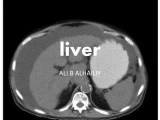
Surface Anatomy Notice the surface anatomy of liver, lying high up in the rib cage therefore risk of injury during chest trauma / pleural tapping etc….
Surface Anatomy Even higher when patient lies down….
Liver surgery • Trisegmentectomy • Right lobe + medial segment of Left lobe • Left lobe + either anterior or posterior segment of Right lobe • Lobectomy • Either entire Right or Left lobe • Segmentectomy • Removal of an anatomical Couinaud’s segment • Non anatomical resection • Wide local excision
Liver Benign Lesions • Cyst • Haemangioma • Focal Nodular Hyperplasia • Hepatic Adenoma • Malignant protential
LiverCysts • Simple cysts • Asymptomatic • Large size – pain • Complications… haemorrhage / abscess • Drainage / Marsupialization of the cyst (lap / open) • Hydatid Cyst • Polycystic Liver Disease • Other organs – spleen / kidneys • Auto Dominant (when associate with kidneys chromosome 16) • Asymptomatic • Complications: • Haemorrhage into cyst • Secondary infection – abscess • Liver dysfunction – totally replaced by cysts… liver transplant
LiverCysts • Pyogenic liver abscess • Streptococcu milleri • Amoebic liver abscess • Entamoeba histolytica • Dysentery • Amoebic cyst ingested – trophozoite form in colon through portal blood into liver • Isolation of the parasite from liver and faeces (microscopy)metronidazole
LiverHydatid Cysts • Echinococcus granulosus (mild) • Echinococcus multilocularis (severe) • Present in dog intestine, ova ingested by human and pass into the portal blood flow • No epithelial lining… fibrous cyst wall • External laminated hilar membrane • Internal enucleated germinal layer • Responsible for the production of colourless hydatid fluid, brood capsule and daughter cysts • 70% Right lobe, 15% Bilateral • Brood capsules • Part of the internal germinal layer • Cellular masses… together with calcerous bodies form the hydatid sand • AXR – calcified cyst wall • CT abdomen • daughter cysts, hydatid sand, laminated cyst wall, floating membrance • Serology • hydatid antigen (ELISA) • Complications • Rupture into peritoneum – may cause anaphylaxis • Rupture into biliary tree – obstructive jaundice, cholangitis • Rupture into pleural cavity – empyema • Rupture into stomach • Rx • Mebendazole +/- surgery • Aspiration + instillation of scolicidal agents not recommended
Liver Haemangioma • Cystically dilated blood filled space with endothelial lining and fibrous septae in between • Female • Symptoms relate to size • Size: pain, rupture • Local pressure: bile duct obstruction / gastric outlet obstruction (very rare) • USG • hyperechoic lesion…can see blood movement inside • CT • progressive enhancement of periphery with central hypodensity remain • Angiogram • cotton wool appearance – rapid filling of the periphery • NO Malignant Potential • Surgery only for very large size with symptom • Since risk of rupture
Focal Nodular Hyperplasia • Consist of normal hepatic cells mixed with bile ductules and divided by firbrous septae… i.e. focal overgrowth of functioning liver tissue supported by fibrous stroma • Benign • Difficult differentiating this from other hepatic lesions... • History & Examination… Tumor markers • CT • Vascular lesion with central scarring • Sulphur colloid liver scan • FNH has kupffer cells and will take up sulphur colloid, but HCC or adenoma lack Kupffer cell therefore will be negative • Biopsy • Close Vs Open… FNA Vs Excisional biopsy
Hepatic Adenoma • Premalignant • Can harbor foci of HCC • Female / Oestrogen (OCP) / Pregnancy • Pale yellow fleshy appearance • 30% multiple • USG • Mixed echogenicity • CT & Angiogram • Hypervascular tumor with irregular hypodense area within • Biopsy • FNAC / Excisional Bx • Mx: Surgical Excision
Malignant Liver Tumor • Primary • Hepatocellular Carcinoma • 85-90% of all primary liver malignant tumour • Cholangiocarcinoma • Angiosarcoma • Hepatoblastoma • Secondaries • Colon • Breast • Lung • etc….
Hepatocellular Carcinomasome histological subtypes • Fibrolamellar carcinoma • Younger patients without cirrhosis • Better prognosis • Adenomatous hyperplasia • Premalignant lesion • Otherwise histologic variations are of little importance • No survival difference
HCC • Spread • Local extension • Diaphragm • Vascular encasement • Biliary obstruction • Portal and Hepatic veins • Rupture --- arterial bleed • Distant Metastasis • Bone • Adrenal glands • Lungs • Brain
HCC • Mortality index 0.94 • Hepatitis B • Presence in 13-73% of all HCC patient • Hepatitis C • Presence in 11-88% of all HCC patient • Cirrhosis • ETOH related • From metabolic disorders • Haemochromatosis • Wilson’s disease • Alpha 1 antityrpsin deficiency • etc… • Aflatoxins
HCC • Late presentation • Thus poor prognosis, high mortality index • Non specific symptoms - late presentation • Pain • Abdominal distention • Weight lost • Dyspepsia • Epigastric mass • Ascites • GIB • Rupture HCC
More tendency to rupture then other tumor Arterial supply tumor Superficial location in liver Tend to seed and invade portal venous system….engorged arterial system Central necrosis….rapid growth Management for rupture HCC Transarterial embolisation Contraindication: portal vein thrombosis Hepatic artery ligation (open surgery) Local tumor plication / resection Surgical packing HCCRupture
Post embolization syndrome • Fever • Abdominal pain • Nausea • Raised serum transaminase • Hepatic abscess • Cholecystitis / gallbladder infarction • Emboli particles get into cystic artery • Upper GIB • Gastric / duodenal wall necrosis • Emboli particles get into right gastric / gastroduodenal artery • Acute pancreatitis • Result from ischaemic assault (or chemo agent irritation if in case of TACE ) during emobilization • Rupture of the tumor – from tumor necrosis • PPI should be routinely used • Check serum amylase • Ultrasound / CT if clinical suspicious
HCC Investigations • History – risk factors / symptoms • Examination • chronic liver disease, complications, masses • LFT … ALP first to derange • Tumor markers AFP • USG • CT • Three phase CT scan • Angiogram • Lipiodol • Oily derivative of poppy seed bind with iodine contrast
AFP • Most widely used… lack sensitivity • Normal 40-50% of HCC patients • Chronic active hepatitis 30% • Diagnostic if >500ng/mL • Single chain polypeptide • Oncofetal antigens • Marker for • HCC & • non seminomatous testicular tumor Alternative for HCC: Des-gamma-carboxy-prothrombin (more specific)
HCC Investigations • USG abdomen • Site, size • Portal vein patency • Biliary tree status • Ascites • CT abdomen • Triphasic CT abdomen • Arterial phase – show hypervascular tumor • HCC • Metastatic Neuroendocrine tumor • Portal dominant phase – show hypovascular tumor • Metastasis • Cholangiocarcioma
HCC Investigations • Lipiodol • Tumor lack kupffer cells therefore unable to remove lipiodol… • Inject into tumor feeding vessel during angiogram, usually with embolisation as well….to decrease tumor vascularity • Plain CT abdomen 10 days after injection of lipiodol • Hepatic Angiogram (HAG) • Arteriovenous fistula • Lipiodol will not stay therefore negative uptake…it all get shunted away • Also will have problem with TAC
HCCManagement • Curative Vs Palliative intent • Assess the clinical stage of the disease • Assess the patient liver status / function • Assess the tumor anatomical resectability
HCCTreatment / Therapy • Surgical Resection • Local Ablative Therapies • Ethanol injection • RFA radiofrequency ablation • Cryosurgery • Laser ablation • Chemotherapy • Local Chemotherapy • TACE • transarterial chemo-embolization • TOCE • transarterial oily chemo-embolization • Intraarterial infusion • surgically placed arterial catheter, need to ligate other branches… right gastric, cholecystectomy & cystic artery ligation… before starting the infusion • Systemic Chemotherapy • Multimodality Therapy • Chemoirradiation
HCC Resectability • Resectability of the tumor depends on two main factors • Disease • Multifocal / bi lobe disease • Extrahepatic disease • Portal vein / IVC / major vascular involvement • Liver status • Hepatic function • Liver volume
Hepatic Function • Child-Turcotte-Pugh Classification • Glucose tolerance tests • Redux tolerance index • Indocyanine green clearance • After 15 mins, >15-20% retention then not for major liver resection
Child-Turcotte-Pugh Classification • A 5-6 • B 7-9 • C 10-15
Liver Volume Assessment • CT volumetric analysis • Anticipated liver remnant volume • The volume of liver remain after surgery… need to have adequate volume to sustain life • Normal liver at least >25% • Disease liver at least >40% • Portal vein embolization (PVE) • When the anticipated liver remnant volume is not enough, just slightly short of requirement • Induce hypertrophy of the future liver remnant • Wait 4 wks for reassessment after embolization
Risk of Local Recurrence • Resection margin … • Not only clearance, but distance of margin as well >1cm better • Size of tumor • No. of tumor • Cirrhosis • Hep B / C status • Positive serology 5 years disease free survival 26-38% • Negative serology 5 years disease free survival 79% • Capsular involvement • Vascular involvement • Lack of Pseudocapsule • Location of tumor • Deep seated tumor… higher recurrent risk • High histological grade
Liver metastasisSecondaries • Aggressive approach • Exclude extrahepatic disease before consider surgery • Single met lesion • 5 years survival 35% • 5 years disease free survival 25% • Multiple met lesions • 5 years survival 25-37% • 5 years disease free survival 20-30% • Repeated resection for recurrent liver met • 5 years survival • After first resection 43% • After second resection 22%
Liver metastasisCa colon • Ca colon – main source • 40% recurrence in liver alone • Poor prognostic factors: • Disease free (ca colon) interval: <1 year worst • Old age • Unclear initial resection margin for the primary tumor (colon) • Advance stage of the primary tumor • Right side colon • Extrahepatic disease (esp. unresectable) • No. of hepatic recurrence (>4 worst) • Size of the largest metastatic lesion (tumor load) • CEA level • Resection margin for the liver metastatic lesion • Segmental Vs non anatomical resection • Absence of capsule / microscopic fibrous pseudocapsule for the metastatic lesion