
Liver
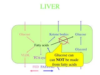
Liver. Function: 1-Metabolic : Glucose 2-Synthetic : Albumin, clotting factors ….. 3-Detoxification : Drugs, hormones , NH3 4-Storage : Glycogen, TG, Fe, Cu, vit 5-Excretory : Bile. Net wt. 1400 – 1600gm (2.5% of body wt)

Liver
E N D
Presentation Transcript
Function: 1-Metabolic : Glucose 2-Synthetic : Albumin, clotting factors ….. 3-Detoxification : Drugs, hormones , NH3 4-Storage : Glycogen, TG, Fe, Cu, vit 5-Excretory : Bile
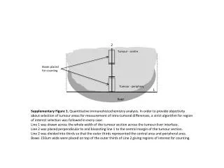
Net wt. 1400 – 1600gm (2.5% of body wt) - Blood supply: Portal v : 60 - 70% Hepatic a : 30 - 40% • Microstructure • Hexagonal lobules →6 acini • Acinus is divided into 3 zones: 1-Zone 1 Periportal areas – closet to the vascular supply 2-Zone 3 Pericentral area 3-Zone 2 Inrermediate bet. Zone 1&2
The parenbchyma is organized into plates of hepatocytes Hepatocytes are radially oriented around terminal hepatic vein ( central v.) -Hepatocytes show only minimal variation in the overall size but nuclei may vary in size , number & ploidy esp. with advancing age -Vascular sinusoids present bet. cords of hepatocytes
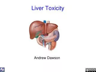
Hepatic injury 1-Inflammation (Hepatitis) 2-Degeneration : ballooning degeneration feathery degeneration:retained biliary material accumulation of iron ,copper
3-Steatosis ( fatty change) microvesicular: ALD Reye syndrome acute fatty change of pregnancy macrovesicular: DM obesity
4-Necrosis -Depending on the type: Coagulative necrosis Councilman bodies Lytic necrosis - Depending on the cause Ischemic Toxic
- Depending on location Centrilobular necrosis: Mid zonal : Periportal : interface hepatitis Focal: Piece meal necrosis bridging necrosis Diffuse: massive & submassive necrosis
5-Regeneration -evidenced by increased mitosis or cell cycle markers. -the cells of the canal of Hering are the progenitor for hepatocytes & bile duct cells (oval cells ).
6-Fibrosis bridging fibrosis 7-Cirrhosis micronodular Macronodular 8-Ductular proliferation
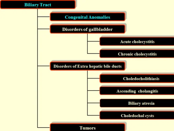
CLINICAL SYNDROMES • The major clinical syndromes of liver disease are: • 1-hepatic failure • 2-cirrhosis • 3-portal hypertension • 4-cholestasis.
liver failure • The alterations that cause liver failure fall into 3 categories: • 1- Acute liver failure with massive hepatic necrosis • 2- Chronic liver disease • 3- Hepatic dysfunction without overt necrosis.
1-Acute liver failure. • This is most often caused by drugsor fulminant viral hepatitis. • Acute liver failure denotes clinical hepatic insufficiency that progresses from onset of symptoms to hepatic encephalopathy within 2 to 3 weeks. • A course extending as long as 3 months is called subacute failure.
The histologic correlate of acute liver failure is massive hepatic necrosis. • It is an uncommon but life-threatening condition that often requires liver transplantation.
2-Chronic liver disease • This is the most common route to hepatic failure and is the end point of relentless chronic liver damage ending in cirrhosis.
3-Hepatic dysfunction without overt necrosis. • Hepatocytes may be viable but unable to perform normal metabolic function: • 1- Acute fatty liver of pregnancy (which can lead to acute liver failure a few days after onset) • 2- Tetracycline toxicity • 3- Reye syndrome
Clinical features 1-Jaundice 2-Hypoalbuminemia →edema 3-Hyperammonemia 4-hyperestrogenemia 5-Spider angiomas 6-Hypogonadism & gynecomastia
Complications: 1-Multiple organ failure e.g lung 2-Coagulopathy → bleeding def. factors II, VII, IX, X 3-Hepatic encephalopathy 4-Hepatorenal Syndrome
Alcoholic liver disease -Alcohol is most widely abused agent -Excessive ethanol consumption causes more than 60% of chronic liver disease in most Western countries and accounts for 40-50% of deaths due to cirrhosis. -It is the 5th leading cause of death in USA due to : 1.Accident 2.Cirrhosis
Pathogenesis • Short-term ingestion of as much as 80 gm of ethanol/d (8 beers or 7 ounces of 80-proof liquor) generally produces mild reversible hepatic changes. • Chronic intake of 50 to 60 gm/day is considered a borderline risk for severe injury. • Women seem to be more susceptible to hepatic injury than are men because of low gastric metabolism of ethanol and differences in body composition.
-80–100 mg/dl is the legal definition for driving under the influence of alcohol -44 ml of ethanol is required to produce this level in 70kg person -In occasional drinkers, bl. Level of 200 mg/dl produces coma & death & resp. failure at 300-400 mg/dl
Habitual drinkers can tolerate levels up to 700 mg/dl without clinical effect due to metabolic tolerance explained by 5-10X induction of cytochrome P-450 system that includes enzyme CYP2E1 which increases the metabolism of ethanol as well as other drugs as cocaine & acetominophen .
Forms of alcoholic liver disease 1-Hepatic steatosis (90-100% of drinkers) 2-Alcoholic hepatitis ( 1- 35% of drinkers) 3-Cirrhosis ( 14% of drinkers) • Steatosis & hepatitis may develop independently
Hepatic steatosis -Can occur following even moderate intake of alcohol in form of microvesicular steatosis • initially centrilobular but in severe cases it may involve the entire lobule . -Chronic intake → diffuse steatosis -Liver is large ( 4 – 6 kg) soft yellow & greasy -Continued intake →fibrosis -Fatty change is reversible with complete absention from further intake of alcohol
Alcoholic hepatitis Characteristic findings : 1-Hepatocyte swelling & necrosis -Accumulation of fat & water & proteins -Cholestasis -Hemosiderin deposition in hepatocytocytes & kupffer cells 2-Mallory-hayline bodies -eosinoplilic cytoplasmic inclusions in degenerating hepatocytes formed of cytokeratin infermediate filaments & other proteins
- Mallory-hayline inclusions are characteristic but not pathognomonic of alcoholic liver disease, they are also seen in : 1-Primary biliary cirrhosis 2-Wilson disease 3-Chronic cholestatic syndromes 4-Hepatocellular carcinoma
3-Neutrophilic reaction 4-Fibrosis -Sinusoidal & perivenular fibrosis -Periportal fibrosis 5-Cholestasis 6-Mild deposition of hemosiderin in hepatocytes & kupffer cells
Alcoholic cirrhosis -Usually it develops slowly -Initially the liver is enlarged yellow but over years it becomes brown shrunken non-fatty organ s.t < l kg in wt. -Micronodular → mixed micro & macronodular -Laennec cirrhosis = scar tissue -Bile stasis -Mallory bodies are only rarely evident at this stage -Irreversible -It can develop rapidly in the presence of alcoholic hepatitis (within 1-2 yrs).
Ethanol metabolism Ethanol → acetaldehyde CH3 CH2OH CH3 C=O H ↑ -Alcohol dehydrogenase (stomach + liver) -Cytochrome P-450 -Catalase ( liver) -
Acetaldehyde → Acetic acid ↑ Aldehyde dehydrogenase
After absorption ethanol is distributed as Acetic acid in all tissues & fluid in direct proportion to blood level • Women have lower levels of gastric alcohol dehydrogenase activity than men & they may develop higher blood Levels than men after drinking the same quantity of ethanol.
- Less than 10% of absorbed ethanol is excreted unchanged in urine , sweat & breathe -There is genetic polymorphism in aldehyde dehydrogenase that affect ethanol metabolism e.g 50% of chinese , vietnamase & Japanese have lowered enzyme activity due to point mutation of the enzyme. → accumulation of acetaldehyde → facial flushing, tachycardia & hyperventilation. • -
Mechanism of ethanol toxicity 1-Fatty change a-Shunting of lipid catabolism toward lipid bio-synthesis due to excess production of NADH over NAD in cystol & mitochondria b-Acetaldehyde forms adducts with tubulin & ↓ function of microtubules → ↓ in lipoprotein transport from liver c- ↑ peripheral catabolism of fat → ↑ FFA delivery to the liver d- ↓ sec. of lipoproteins from hepatocytes e. ↓ oxidation of FFA by mitochondria 2-Induction of cytochrome P-450 enhances the metabolism of drugs to toxic metabolites (e.g acetominophen )
3. ↑free radicals production due to activation of cytochrome P-4so leads to membrane & protein damage 4. Alcohol directly affect microtubular & mitochondrial function & membrane fluidity 5.Acetaldehyde causes lipid peroxidation & antigenic alteration of hepatocytes → immune attack 6. Superimposed HCV infection causes acceleration of liver injury (HCV hepatitis occurs in 30% of alcoholics )
7. Alcohol → release of bacterial endotoxins into portal circulation from the gut → inflammation of the liver 8. Alcohol → regional hypoxia in the liver due to release of endothelins which are potent vasoconstrictors → ↓ hepatic sinusoidal perfusion 9. Alteration of cytokine regulation TNF is a major effector of injury IL6 IL8 IL18
Clinical features -Hepatic steatosis ( reversible ) • ↑ liver • ↑ liver enz. • Severe hepatic dysfunction is unusual -Alcoholic hepatitis • 15-20 yr. of excessive drinking • Non-specific symptoms, malaise, anorexia, wt. loss • Hepatosplenomegaly • ↑ LFT Each bout of hepatitis →10-20% risk of death → cirrhosis in 1/3 in few yrs. -Cirrhosis Portal hypertension
Causes of death in alcoholic liver disease: 1-hepatic failure 2-Massive GI bleeding 3-Infections 4-Hepatorenal syndrome 5-HCC in 3-6% of cases


![[EPUB] DOWNLOAD Natural Liver Cleanse Recipes: Liver cleanse juices, liver cleanse tea, Liver cleanse soup, fatty liver](https://cdn7.slideserve.com/12507667/slide1-dt.jpg)