
Liver
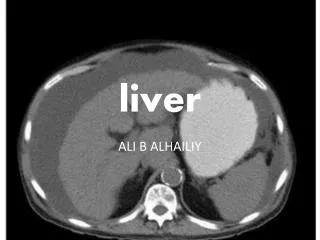
Liver. Basic facts. Lies mainly in right hypochondrium and epigastrium , extending into part of left hypochondrium Increases in size rapidly, achieving maximum size at the age of 18 Gradual decrease in size from middle age onwards. Weight – infants 5% of total body weight adults- 2%

Liver
E N D
Presentation Transcript
Basic facts • Lies mainly in right hypochondrium and epigastrium, extending into part of left hypochondrium • Increases in size rapidly, achieving maximum size at the age of 18 • Gradual decrease in size from middle age onwards
Weight – infants 5% of total body weight adults- 2% • Wedge shaped- shape determined by the form of upper abdominal cavity into which it grows • Reddish brown colour – varies according to fat content
Obesity is commonest factor leading to increase in fat content [steatosis] • Increased fat – yellow tinge • Soft to firm consistency – partly dependant on blood volume in liver and fat content
Surfaces • Diaphragmatic • Visceral
Superiorly • Right dome of diaphragm • Heart • Left dome of diaphragm [partly]
Anteriorly • Anterior attachment of diaphragm • Right pleura • Right 6th- 1oth ribs and their costal cartilages • Left 7th and 8th costal cartilages
Right • Right dome of diaphragm • Right lung and basal pleura • Costophrenic recess • 9th and 10th ribs
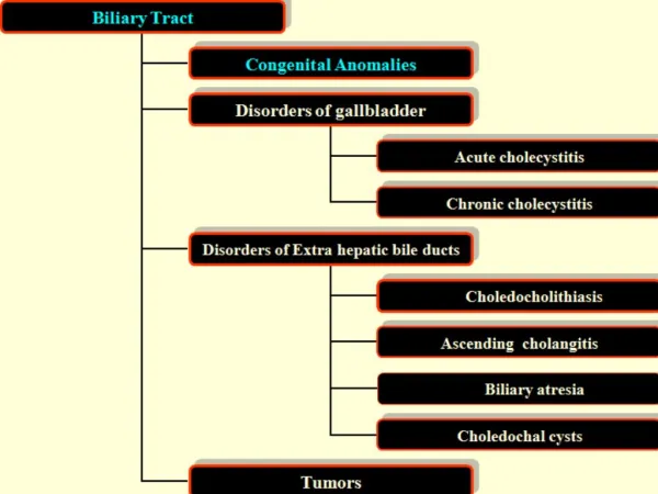
Relations • Gall bladder • Oesophagus • Fundus of stomach • Pylorus • 1st part of duodenum
Hepatic flexure of colon • Right suprarenal gland • Right kidney `
Blood supply • Dual- • venous blood via portal vein and arterial supply by hepatic artery: • both vessels enter the liver at the hilum or portahepatis • 75-80% of blood supply to the liver comes from portal vein, which carries oxygen-poor blood from digestive tract, pancreas and spleen
The portal blood carries • Nutrients and toxic materials absorbed in the intestine • Blood cells and breakdown products of blood cells from the spleen • Endocrine secretions of the pancreas and enteroendocrine cells of GIT
The hepatic artery carries oxygenated blood from the liver, providing 20-25% of its blood supply • Blood from 2 sources gets mixed before it perfuses the hepatocytes • The distributing branches of portal vein and hepatic artery supply the sinusoidal capillaries that bathe the hepatocytes
The sinusoids are in intimate contact with the hepatocytes and provide for exchange of substances between blood and liver cells • The sinusoids lead to a central vein, that in turn opens into the sublobular veins • Blood leaves the liver through hepatic veins, which empty into the IVC


![[EPUB] DOWNLOAD Natural Liver Cleanse Recipes: Liver cleanse juices, liver cleanse tea, Liver cleanse soup, fatty liver](https://cdn7.slideserve.com/12507667/slide1-dt.jpg)