Download
1 / 16
160 likes | 473 Vues
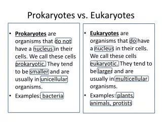
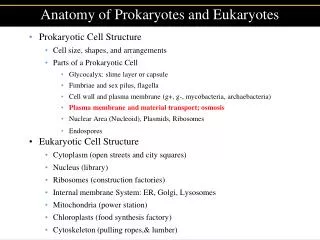
Anatomy of Prokaryotes and Eukaryotes. Prokaryotic Cell Structure Cell size, shapes, and arrangements Parts of a Prokaryotic Cell Glycocalyx: slime layer or capsule Fimbriae and sex pilus, flagella Cell wall and plasma membrane (g+, g-, mycobacteria, archaebacteria)

E N D
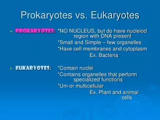
Anatomy of Prokaryotes and Eukaryotes • Prokaryotic Cell Structure • Cell size, shapes, and arrangements • Parts of a Prokaryotic Cell • Glycocalyx: slime layer or capsule • Fimbriae and sex pilus, flagella • Cell wall and plasma membrane (g+, g-, mycobacteria, archaebacteria) • Plasma membrane and material transport; osmosis • Nuclear Area (Nucleoid), Plasmids, Ribosomes • Endospores • Eukaryotic Cell Structure • Cytoplasm (open streets and city squares) • Nucleus (library) • Ribosomes (construction factories) • Internal membrane System: ER, Golgi, Lysosomes • Mitochondria (power station) • Chloroplasts (food synthesis factory) • Cytoskeleton (pulling ropes,& lumber)
Plasma or Cell Membrane Figure 4.14a
Types of Transport Across a Selectively Permeable Cell Membrane
Osmosis and Water Balance in Cells Osmosis is the passive transport of water across a selectively permeable membrane. • Water moves across a membrane from high concentration (high purity) to low concentration (low purity)
Osmosis: Water Moving From High Purity to Low Purity (outside) (outside) Bacterial growth media is made to be isotonic with cells ( ~ 1% dissolved substances) Figure 4.18c-e
Anatomy of Prokaryotes and Eukaryotes • Prokaryotic Cell Structure • Cell size, shapes, and arrangements • Parts of a Prokaryotic Cell • Glycocalyx: slime layer or capsule • Fimbriae and sex pilus, flagella • Cell wall and plasma membrane (g+, g-, mycobacteria, archaebacteria) • Plasma membrane and material transport; osmosis • Nuclear Area (Nucleoid), Plasmids, Ribosomes • Endospores, Cytoskeleton • Eukaryotic Cell Structure • Cytoplasm (open streets and city squares) • Nucleus (library) • Ribosomes (construction factories) • Internal membrane System: ER, Golgi, Lysosomes • Mitochondria (power station) • Chloroplasts (food synthesis factory) • Cytoskeleton (pulling ropes,& lumber)
Inclusions and Other Membranes Inclusion Nucleoid (Nuclear Area) Plasmid Figure 4.6a
Ribosomes Ribosomes from eukaryotes are larger and constructed differently Figure 4.19
Intracellular Membranes and Inclusions • Metachromatic granules (phosophate) Polysaccharide granules • Lipid inclusions • Sulfur granules • Carboxysomes • Thylakoids/photsynthetic membranes • Gas vacuoles • Magnetosomes
Endospore Formation Endospores are heat-resistant, UV resistant, and chemically resistant Figure 4.21a
Bacterial Cell Walls Include Scaffolding Proteins (Cytoskeleton) Proteins similar to actin in eukaryotes help in expanding the peptidoglycan during bacterial growth by acting as scaffolds
Anatomy of Prokaryotes and Eukaryotes II • Prokaryotic Cell Structure • Cell size, shapes, and arrangements • Parts of a Prokaryotic Cell • Glycocalyx: slime layer or capsule • Fimbriae and sex pilus, flagella • Cell wall and plasma membrane (g+, g-, mycobacteria, archaebacteria) • Plasma membrane and material transport; osmosis • Nuclear Area (Nucleoid), Plasmids, Ribosomes • Endospores, Cytoskeleton • Eukaryotic Cell Structure • Cytoplasm (open streets and city squares) • Nucleus (library) • Ribosomes (construction factories) • Internal membrane System: ER, Golgi, Lysosomes (Construction & shipping) • Mitochondria (power station) • Chloroplasts (food synthesis factory) • Cytoskeleton (pulling ropes,& lumber)
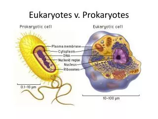
Greater Size, Complexity, and Nucleus in Eukaryotes Figure 4.22b
Eukaryotic Cells:Protists, Plants, Animals, & Fungi Cell wall (if present) Figure 4.21a
Anatomy of Prokaryotes and Eukaryotes • Prokaryotic Cell Structure • Cell size, shapes, and arrangements • Parts of a Prokaryotic Cell • Glycocalyx: slime layer or capsule • Fimbriae and sex pilus, flagella • Cell wall and plasma membrane (g+, g-, mycobacteria, archaebacteria) • Plasma membrane and material transport; osmosis • Nuclear Area (Nucleoid), Plasmids, Ribosomes • Endospores • Eukaryotic Cell Structure • Cytoplasm (open streets and city squares) • Nucleus (library) • Ribosomes (construction factories) • Internal membrane System: ER, Golgi, Lysosomes • Mitochondria (power station) • Chloroplasts (food synthesis factory) • Cytoskeleton (pulling ropes,& lumber)