Chapter 10: Gram-Positive Rods
Chapter 10: Gram-Positive Rods. Corynebacterium (Coryneforms) Genus: Corynebacterium Species: Corynebacterium diphtheriae Corynebacterium xerosis Corynebacterium pseudodipheriticum

Chapter 10: Gram-Positive Rods
E N D
Presentation Transcript
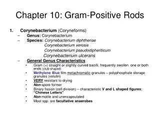
Chapter 10: Gram-Positive Rods • Corynebacterium (Coryneforms) • Genus: Corynebacterium • Species: Corynebacterium diphtheriae Corynebacterium xerosis Corynebacterium pseudodipheriticum Corynebacterium ulcerans • General Genus Characteristics • Gram (+) straight or slightly curved bacilli, frequently swollen one or both ends (club-shaped) • Methylene Blue filmmetachromatic granules – polyphosphate storage granules (velutin) • VERY resistant to drying • Non-spore former • Binary fission (cell division) – characteristic V and L shaped figures; “Chinese Letters” • Non-motile and unencapsulated • Most spp. are facultative anaerobes
كرينه باكتريوم • محيطهاي كشت : • تلوريت پتاسيم • لفلر • تينسدال • محيط پايهٌ قندي CTA
كرينه باكتريوم • بيوتايپهاي كرينه باكتريوم ديفتريه در محيط تلوريت پتاسيم از هم جدا مي شوند : • بيوتايپ گراويس به شكل گل مينا و بزرگ • بيوتايپ مي تيس به اشكال مختلف(تخم مرغ نيمرو)و متوسط • بيوتايپ اينتر مديوس كوچكتر از بقيه و به شكل تخم قورباغه
Gram-Positive Rods • Corynebacterium (Coryneforms) • Growth Conditions • Selective Media – blood tellurite reduction of tellurite gray/black colonies • 3 biotypes of C. diphtheriae • Gravis: grave infection • Intermidius: intermediate infection • Mitis: little infection ***All 3 serotypes are capable of producing the same toxin & clinical disease.
Gram-Positive Rods • Corynebacterium (Coryneforms) • Corynebacterium diphtheriae • Normal Flora – URT (throat and Nasopharynx) • Reservoir – Human pathogen ONLY • Infection • Respiratory (diphtheria) • Cutaneous (skin infection) • Transmission/Epidemiology – person-to-person (asymptomatic or diseased patients) • Inhalation of aerosols (i.e., respiratory droplets) • Direct contact w/ cutaneous lesions (analogous to impetigo) • Direct contact w/ contaminated fomites=لوازم اشياء (least frequent)
Gram-Positive Rods • Corynebacterium (Corniforms) • Corynebacterium diphtheriae • Virulence Factor • Diphtheria Toxin – very powerful exotoxin • Avirulent C. diphtheriae infected by bacteriophage (tox +) Lysogenic conversion to Virulent stain C. diphtheriae(tox +) Recall:Lysogeny – bacteriophage has ability to insert their DNA into DNA molecule of host bacterium. • Expression of tox gene • Regulated by Fe concentration • Tox gene repressor: Fe-containing protein • Function of repressor – inhibits toxin production • [Fe2+] decreased limited repressor available toxin gene
Gram-Positive Rods • Corynebacterium (Corniforms) • Corynebacterium diphtheriae • Pathogensis • Diphtheria Toxin – 2 subunit fragments A & B • B Fragment: Binds to specific cell surface receptors Mediates transport of A fragment into cell • A Fragment: Inhibits protein synthesis => cell death/necrosis • Specifically, fragment A catalyzes a rxn. between NAD+ and the polypeptide chain elongation factor, EF-2: transfer of ADPR from NAD+ to EF-2 • Result = ADPR:EF-2 complex is INACTIVATED & protein (peptide) synthesis is terminated
Gram-Positive Rods • Corynebacterium (Corniforms) • Corynebacterium diphtheriae • Clinical Disease • Localized infection • Respiratory (Pharyngeal Diphtheria) • Cutaneous Diphtheria (both toxigenic & non-toxigenic strains) • Systemic infection - toxemia
Gram-Positive Rods • Corynebacterium (Corniforms) • Corynebacterium diphtheriae • Pharyngeal Diphtheria Mode of Infection • Inhalation • Attachment – to mucous membranes of passageways of oral cavity • Attachment/Colonization – non-specific virulence factors • Toxin production – necrosis of mucosal cells lesion expansion=توسعه بسط انبساط • Pseudomembrane formation – grayish-white patches=تكه وصله مشمع روي زخم of thick fibrous exudate (neutrophils, corynebacterium, epthelial cells) • Tight=سفت محكم مانع دخول هوا adherence – to epithelial surface w/ no invasion of underlying tissue • Spread of pseudomembrane – involvement of nasopharyngeal tissues (mouth, pharynx) or larynx & trachea • Entry of toxin into the blood stream – multi-organ involvement: heart, CNS and kidneys; most frequent & serious damage d/t toxin occurs in the heart and CNS, w/ death resulting from damage to the heart.
Gram-Positive Rods • Corynebacterium (Corineforms) • Corynebacterium diphtheriae • Pharyngeal Diphtheria: Summary • Location infection of the throat (URT infection) • Production of thick, grayish, adherent exudate • Pseudomembrane • Composition = cell debris from mucosa & inflammatory products • Coats the throat; may extend into nasal passages or downward into RT, obstructing the airways leading to suffocation • w/ disease progression – generalized sx’s d/t production & absorption of toxin • MAJOR CLINICAL EFFECTS: Heart & Peripheral Nerve involvement • Cardiac conduction defects & myocarditis CHF & permanent heart damage. • Neuritis of CN’s & paralysis of muscle grps – seen in late dis.
Gram-Positive Rods • Corynebacterium (Corineforms) • Corynebacterium diphtheriae • Cutaneous Diphtheria • Localized infections arising from: insect bites, skin trauma, poor personal hygiene • Ex. Puncture wound or cut in skin introduction of bacterium into subcutaneous tissue chronic, non-healing ulcer w/ grayish membrane. • Risk Group: homeless • Sx’s: impetigo-like lesions
Gram-Positive Rods • Corynebacterium (Corineforms) • Corynebacterium diphtheriae • Systemic Spread of Primary Infection • Heart – myocarditis • CNS – cranial & peripheral; nerve degeneration (paralysis) • Kidneys – filtering inadequacies; kidney failure • Risk Groups • Individuals w/ low socioeconomic status • Crowded & unsanitary conditions • Non-immunized children (1-10 yoa) • Elderly – low immunization status
Gram-Positive Rods • Corynebacterium (Corineforms) • Corynebacterium diphtheriae • Diagnosis • Clinical: sx presentation (gray-membrane w/ swelling) • Serological: Ab detection (to the toxin Ag.) • Laboratory identification • Specimen – swab cultures ( pharynx, nasopharynx, or cutaneous sites) • Microscopic appearance – smears alone are unreliable, possible presence of diphtheriods • Cultivation – Media • Loeffler media Overnight growth film (Methylene Blue) metachromatic granules • Selective media (Tinsdale agar, containing tellurite) Blood tellurite corynebacterium reduce tellurite gray/black colored colonies w/ halos Tinsdale’s Agar contains Potassium tellurite, an inhibitor of other respiratory flora
Gram-Positive Rods • Corynebacterium (Corniforms) • Corynebacterium diphtheriae • Treatment: • Respiratory Diphtheria • Passive Immunization: DAT (Diphtheria antitoxin) => for neutralization of the toxin • Antibiotic Tx: penicillin or erythromycin => slows infection spread by killing the organism, preventing further toxin production • Cutaneous Diphtheria • Compresses soaked in penicillin applied to lesions • Control/Prevention • Active Immunization – toxoid vaccine (DPT) • Start @ infancy; booster injections – administered @ ~10yr. Intervals throughout life • Harvest bacteria purify toxin inactive toxin Formaldehyde toxoid
Gram-Positive Rods • Corynebacterium (Corineforms) • Diphtheroids • Other corynebacterium spp. that morphologically resemble C. diphtheriae • Non-pathogenic • Normal flora – eyes (conjunctiva), skin, nose, mouth/throat, vagina and urethra (UT) • NO toxin production • Opportunistic infections, esp. in immunosuppressed pts. • Ex. 1. Corynebacterium xerosis – opportunist in eye & post- operative infections 2. Corynebacterium pseudodipheriticum – endocarditis in pts. undergoing cardiac surgery/valvular prostheses
Gram-Positive Rods • Bacillus • Genus: Bacillus • Species: Bacillus anthracis – causes anthrax Bacillus cereus – GI infection • Microscopic Morphological Appearance • Large, Gram (+) bacillus • ***Spore former*** (Endospores) • Non-motile • Encapsulated • Aerobe (strict or facultative) • Note: most species of Bacillus are found in soil & water, and are usually encounters in the medical laboratory as airborne contaminants • Most clinically important = B. anthracis (Anthrax)
Gram-Positive Rods • Bacillus • Virulence factors • Capsule • Exotoxin – complex of 3 protein factors • Edema Factor (EF) • Protective Agent (PA) • Lethal Factor (LF) => EF + host cell calmodulin • Action of Exotoxin • PA inserts into the cytoplasmic (phospholipid) membrane of host EF binds to PA transfer of EF into host cytoplasm • PA facilitates transfer of EF into host cytoplasm
Gram-Positive Rods • Bacillus • Action of Exotoxin (Pathogenesis) • Rxn: EF + calmodulin → → activation of Adenyl Cyclase [ ATP → cAMP] → ↑cAMP => EDEMA • Increased levels of cAMP → cell oversecretion (EDEMA) • Transmission – animal to man • Cutaneous infection • Pulmonary infection 3 diff. types of infection • Intestinal infection -based upon route of entry
Gram-Positive Rods • Bacillus • Human Anthrax • Causative agent = B. anthacis; rare in US • 1984-1997: only 3 cases of cutaneous anthrax reported • 2001: 20 cases – probable exposure to B. anthracispowder sent thru US mail; unknown source. • Epidemiology • Anthrax is an enzootic disease of worldwide occurrence. • Enzootic diseases are endemic to an animal population; occurrence changes little over time. • Anthrax affects domestic herbivores • Sheep, goats, horses • Transmission to humans by contact w/ infected an. products or contaminated dust (fig 10.6 p. 95)
Gram-Positive Rods • Bacillus • Human Anthrax • Epidemiology • Infection initiated by subcutaneous innoculation of spores thru incidental skin abrasions • Also, but less frequently, inhalation of spore-laden dust causes Pulmonary Anthrax • Spores can remain viable for many years in contaminated pastures, bones, wool, hair, hides, or other an. materials. • Highly resistant to physical & chemical agents
Gram-Positive Rods • Bacillus • 3 different types of B. anthracisinfection • Cutaneous Anthrax – m/c; localized infection of skin; Least dangerous • Inoculation of spores → deposit beneath the skin, where they germinate → resulting in exotoxin production • Malignant pustule – located on hands, forearms and head • Eschar – black scab (painless) • Possible invasion to regional lymph nodes, then entry into general circulation • Pulmonary Anthrax = Woolsorter’s Disease • Most serious, but least common in US; mortality rate ~100% • Inhalation of spores → deposit in lung alveoli • Characterized by progressive hemorrhagic lymphadenitis • Occupational risk groups; transmission of spore to individuals working w/ infected animals • Sx’s: onset – sudden w/ high fever & respiratory distress (similar to respiratory infection. • Pneumonia is often followed by sepsis and death in untx’ed cases
Gram-Positive Rods • Bacillus • 3 different types of B. anthracisinfection • Gastrointestinal Anthrax • Ingestion of spores → exotoxin production in intestinal tract → necrotic lesions • Results from eating contaminated meat • Immunity = permanent immunity is acquired post infection • Treatment: broad spectrum antibiotics; penicillin is NOT recommended b/c of inducible β-lactamase in B. anthracis • Control/Prevention • Sterilization of wool, hair, etc. prior to the handling of animals • Sacrifice of infected animals • Immunization of animals • Vaccine for workers in high risk occupations – purified toxoid • Post-exposure antibiotic prophylaxis
Gram-Positive Rods • Bacillus • Bacillus cereus • Common inhabitant of rice when spores NOT killed properly • Infection/Disease • Gastroenteritis – Food intoxication: eating cooked, improperly stored rice • Emetic-type gastroenteritis • Virulence factor • Enterotoxins • Sx’s • Gastroenteritis – abdominal pain, diarrhea • Emetic-type gastroenteritis: vomiting • Control/prevention • Refrigeration of food – no germination of spores
Gram-Positive Rods • Listeria • Spp. are short, slender, Gram (+) rods • Non-spore formers • Occur in short chains or diplobacilli • Intracellular parasites • Catalase (+) • “tumbling” motility, as seen by light microscopy in liquid medium @ 25°C • Distinct from catalase (-) streptococcus & non-motile corynebacterium • Grow facultatively
Gram-Positive Rods • Listeria • Epidemiology • Listeria monocytogenes= ONLY spp that infects humans • Listeria spp are widespread in nature among animals • Human infections are usually food-borne • 2-3% processed dairy products (ice cream, cheese) • 20-30% ground meats • Majority of retail poultry • Refrigeration does NOT suppress growth in food • 1-15% of healthy humans are asymptomatic intestinal carriers • Infections are m/c in pregnant females, fetuses/newborns, immunocompromised individuals (i.e., elderly, corticosteroid use) • 200 cases reported/yr. in US w/ 450 deaths & 100 stillbirths
Gram-Positive Rods • Listeria • Pathogenesis of L. monocytogenes (fig 10.8 p.98) • Attaches to & enters a variety of mammalian cells by normal phagocytosis (i.e., macrophages) • Escape from phagocytic vacuole d/t formation of Listeriolysin O, a membrane-damaging toxin. • Growth in host cell cytoplasm, stimulating changes in cell function that facilitate its direct passage from cell-to-cell • Assembly of actin filament tail that pushes the bacterium to the surface of the macrophage • Pseudopod extension forms, facilitating transfer into another phagocyte. • Membrane-degrading phospholipases mediate passage of org into next cell w/ IS avoidance
Gram-Positive Rods • Listeria • L. monocytogenes Clinical Disease (Listeriosis) • M/C = septicemia & meningitis • Focal skin lesions (granulomatous) sometimes seen • Immunocompromised individuals w/ defects in cellular immunity – potential serious infections • Pregnant females in 3rd trimester may have mild “flu-like” illness • This and asymptomatic colonization of vaginal tract – can allow organism to be transmitted to newborn • L. monocytogenes = common cause of newborn meningitis
Gram-Positive Rods • Listeria • Laboratory Dx of L. monocytogenes • Isolation of organism from Blood, CSF • Blood agar – production of small colony, surrounded by narrow zone of β-hemolysis • Distinguishable from streptococci • + motility • Production of catalase • Treatment: antibiotic therapy • Prevention: proper food preparation & handling
Gram-Positive Rods • Miscellaneous Non-spore forming, G(+) Rods • Propionibacterium • Common inhabitants of normal skin • Genus of anaerobic or microaerophilic rods of diphtheroid-like morphology • Rare cases of endocarditis • P. acnes = strict anaerobe; causes acne • Lactobacillus • Commensal flora of human mucous membranes • Produce lactic acid during fermentation • Assist in maintaining acid pH of normal mucous epithelia • Acid production by oral lactobacilli => dental caries • Erysipelothrix rhusiopathiae • Filamentous, Gram (+) rod that causes dis. in animals • Rare skin infection in humans who handle animal products (i.e., butchers, DVM’s, fisherman)
Chapter 11: Neisseriae • Genus Neisseriaeconsists of Gram-negative, aerobic cocci. • Organisms primarily reside on mucosal surfaces • 2 Neisseriae spp are pathogenic for humans • N. gonorrhoeae (gonococcus) • Cuasative agent for gonorrhea • N. meningitidis(meningococcus) • Frequent cause of meningitis • Gonocci & Meningococci • Both are non-motile DIPLOCOCCI; look very similar • Both are pyogenic cocci b/c infections are characterized by production of purulent material comprised largely of WBC’s
Neisseriae • General characteristic of the Genus • Gram-negative Diplococci(kidney-bean shaped) • Small, round, glistening, white-to-cream colored after 24 hr. colonization • Non-hemolytic • Non-motile • Non-spore forming • Oxidase-positive(presence of enz. cytochrome oxidase– for rapid dx) • Fastidious growth requirements • Optimal growth temp = 37°C • Aerobic or microaerophilic, utilizing an oxidative form of metabolism • Require atmosphere of 5% CO2 • Require thiamine and thiamine pyrophosphate for nucleic acid biosynthesis • Produces catalases, using the enzymes for fermentation of certain CHO’s • Highly Autolytic – autolysis causes cell wall breakdown (PG fragments) – highly inflammatory • Highly sensitive to dessication d/t capsular polysaccharide • Laboratory tests for distinguishing pathogenic strains from commensal strains • N. gonorrhoeae => (+) for glucose utilization, (-) for maltose utilization • N. meningitidis => (+) for glucose utilization, (+) for maltose utilization
Neisseriae • NEISSERIAE GONORRHOEAE • One of m/freq’ly reported infectious diseases in US • Causative agent = N. gonorrhoeae, a Gram (-) Diplococcus • Commonly found w/in PMN leukocytes of clinical samples • Transmitted via sexual contact; rarely during delivery thru infected vaginal canal • Survival rate outside body = LOW d/t high sensitivity to drying • Epidemiology: major health problem worldwide
Neisseriae • NEISSERIAE GONORRHOEAE • Structure of Gonococci: Unencapsulated, piliated, non-motile, diplococci • Pili – hairlike surf. Appendages; VIRULENCE Factor! • Composed of protein pillin • Enhance attachment to host epithelial & mucosal cell sufaces • Confers resistance to phagocytosis • Potential to produce genetically different pilin molecules over time – antigenic variation or gene conversion
Neisseriae • NEISSERIAE GONORRHOEAE • Structure of Gonococci: Unencapsulated, piliated, non-motile, diplococci • Lipoligosaccharide (LOS) • w/ shorter, more highly branched, non-repeat O-antigenic side chains that do the LPS’s of other Gram-negative bacteria • Human serum IgM Ab’s are directed toward LOS Ag’s • Other Membrane Proteins (OMPs) • Contribute to the virulence of N. gonorrhoeae • OMP II = “opacity protein” b/c its presence renders gonococcal colonies less translucent; along w/ pili – mediated attachment of organism to host cell.
Neisseriae • NEISSERIAE GONORRHOEAE • Pathogenesis • Pili & OMP II => facilitate adhesion to epithelial cells of urethra, rectum, cervix, pharynx, conjunctiva • Make colonization possible • Production of IgA protease that cleaves human IgA Ab’s – evasion of IS response • Seen w/ BOTH gonococci & meningococci
Neisseriae • NEISSERIAE GONORRHOEAE • Clinical Notes • 4th m/c Sexually Transmitted organism today • Colonization of mucous membrane of GU tract of rectum • Cause localized infection w/ production of pus • May lead to tissue invasion, chronic inflammation & fibrosis • Females > males w/ regard to being asymptomatic carriers – can transmit gonococcal infection
Neisseriae • NEISSERIAE GONORRHOEAE • Clinical Diseases • Genitourinary Tract infections • Acute & easy to Dx in Males • Pt. presents w/ yellow, purulent urethral discharge & painful urination. • Female – infection w/in endocervix (cervicitis) & extends to urethra & vagina • m/c presentation = greenish-yellow discharge; intermenstrual bleeding is possible • May progress to uterus, causing Salpingitis (inflammation of Uterine/Fallopian Tubes), Pelvic Inflammatory Disease (PID): gonococci ascend to Reprod. Tract – affect uterus, ovaries & contiguous structures; destruction of upper reproductive tract mucosa • 20% infertility rate w/ gonoccocal salpingitis d/t tubal scarring
Neisseriae • NEISSERIAE GONORRHOEAE • Clinical Diseases • Rectal Infections (Proctitis) – seen w/ male homosexuals • Sx’s: constipation, painful defecation, purulent discharge • Pharyngitis • Contracted by oral-genital contact • Sx’s: purulent exudate; may mimic mild viral or streptococcal sore throat • Opthalmia Neonatoreum • Infection of the conjunctival sac acquired by newborn during passage thru infected birth canal • Blindness if untreated • Tx: 1% Silver Nitrate on eyes prevents disease or 1% Tetracycline or 0.5% erythromycin
Neisseriae • NEISSERIAE GONORRHOEAE • Clinical Diseases • Disseminated Infection/Bacteremia • Bacteremia is rare d/t limited gonococcal ability to multiply in bloodstream (NOTE: meningococcal multiply rapidly in the blood) • Some gonococcal strain w/ ability to cause bacteremia • Gonococci cross mucosal barrier d/t blood-barrier breakdown; blood culture (+) for Gram (-) Diplococci • Sx’s (Both male & female; m/c in females, though): Fever; painful, purulent arthritis; small pustules on skin scattered on a erythematous base – may develop into necrosis • Gonococcal = m/c cause of septic arthritis in sexually active adults
Neisseriae • NEISSERIAE GONORRHOEAE • Clinical Laboratory Identification • Males: numerous neutrophils (PMN’s) containing Gram (-) diplococci in urethral exudate = provisional Dx – start TX. • Females: a positive gonococcal culture needed to Dx infection • If bacteremia suspected – additional cultures from skin lesions, joint fluid, blood
Neisseriae • NEISSERIAE GONORRHOEAE • Clinical Laboratory Identification • Growth conditions • Growth best under aerobic conditions • Most strains require enhanced CO2 • N. gonnorrhoeae ferments glucose (not maltose, lactose or sucrose) • Oxidase-positive • Mixed growth seen on plain Chocolate Agar medium (non-selective medium) • Pure culture (selective growth) seen w/ Thayer-Martin Chocolate Agar Medium – to isolate gonoccoci
Neisseriae • NEISSERIAE GONORRHOEAE • Clinical laboratory Identification • Selective Media • Thayer-Martin chocolate agar medium contains several antibiotics that suppress the growth of non-pathogenic neisseriae and other normal/abnormal flora • Important medium for cultures obtained from GU tract & rectum • Culture of N. gonorrhoeae on Thayer-Martin agar is the “gold-standard” for DX • Treatment & Prevention (diff to control b/c dis tied to sexual promiscuity) • >20% of gonococcal isolates are resistant to penicillin, tetracycline, cefoxitin, =/or spectinomycin • Penicillin-resistant organisms = Penicillinase-producing N. gonorrhoeae • Tx w/ 3rd generation cephalosporins for uncomplicated infections of urethra, endocervix or rectum • Abstinence, avoiding casual sexual contact, limiting # of sexual partners, taking birth control measures (i.e., condom use)
Neisseriae • NEISSERIA MENINGITIDIS • One of the m/c causes of MENINGITIS • Causative agent =N. meningitidis • Infection can take form of fulminant meningococcemia, w/ intravascular coagulation, circulatory collapse, and potentially fatal shock (w/out meningitis) • Outbreaks common in winter & early spring; favored by close contact • Schools, institutions, military barracks • N. meningitidis – afflicts young, previous well individuals; and can progress to DEATH in hours
Neisseriae • NEISSERIA MENINGITIDIS • Structure of meningococci • Non-motile, Gram (-) Diplococci; always found in pairs • Piliated – pili allow attachment to nasopharyngeal mucosa, where organism it harbored in carriers and those w/ disease. • Encapsulated • Meningococcal polysaccharide capsule = antiphagocytic; most important VIRULENCE factor!
Neisseriae • NEISSERIA MENINGITIDIS • Structure of meningococci • Serogroups14 different capsular polysaccharide types (LOS) • Most infections w/ serogroups A, B, C, W and Y • 90% w/ A, B, and C • Serogroup A => massive epidemics in developing countries • Serogroup B=> most predominant cause of disease and mortality in US • Serotypes • 2nd classification sys: serotyping (1,2,3,….20) • Serological, based upon properties of OMPs and LOS
Neisseriae • NEISSERIA MENINGITIDIS • Pathogenesis • Maintenance of infection conferred by the meningococcal capsule w/ antiphagocytic properties • LOS is released during autolysis & bacteial cell division – toxic effects in disseminated dis. • Production of IgA protease that cleaves human Ab IgA – evasion of the IS response
Neisseriae • NEISSERIA MENINGITIDIS • Clinical Notes • N. meningitidis– initial colonization w/in nasopharynx, causing asymptomatic meningococcal pharyngitis • In young & immunocompromised, organism can gain entry to bloodstream (disseminated dis or bacteremia) meningitis +/or fulminant septicemia • N. meningitidis = m/c cause of meningitis